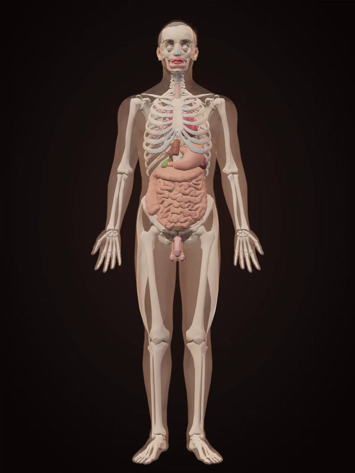
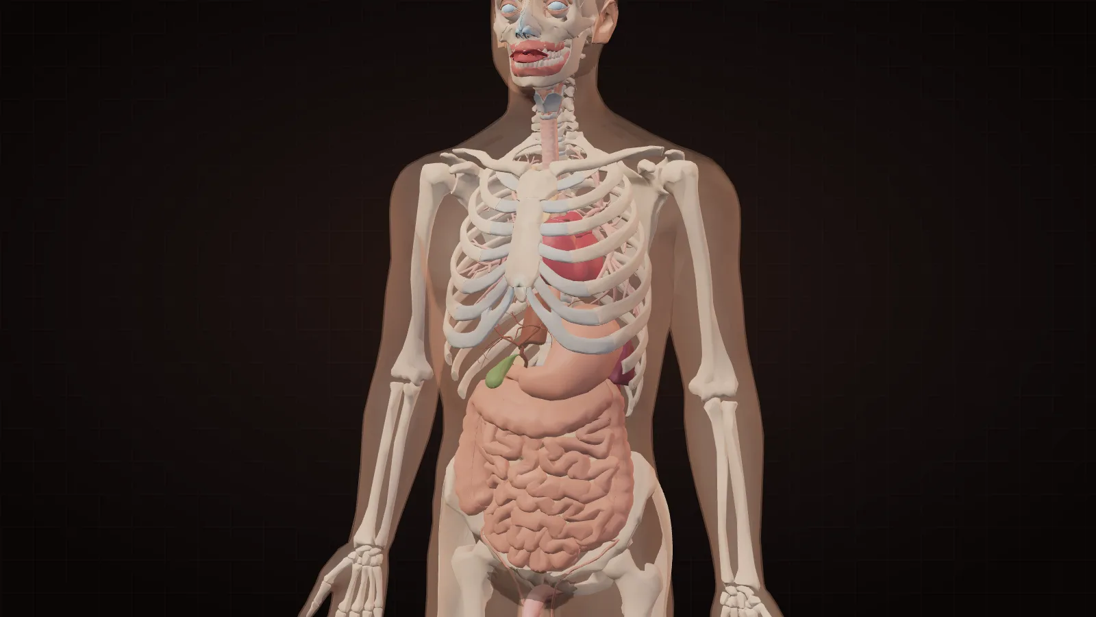
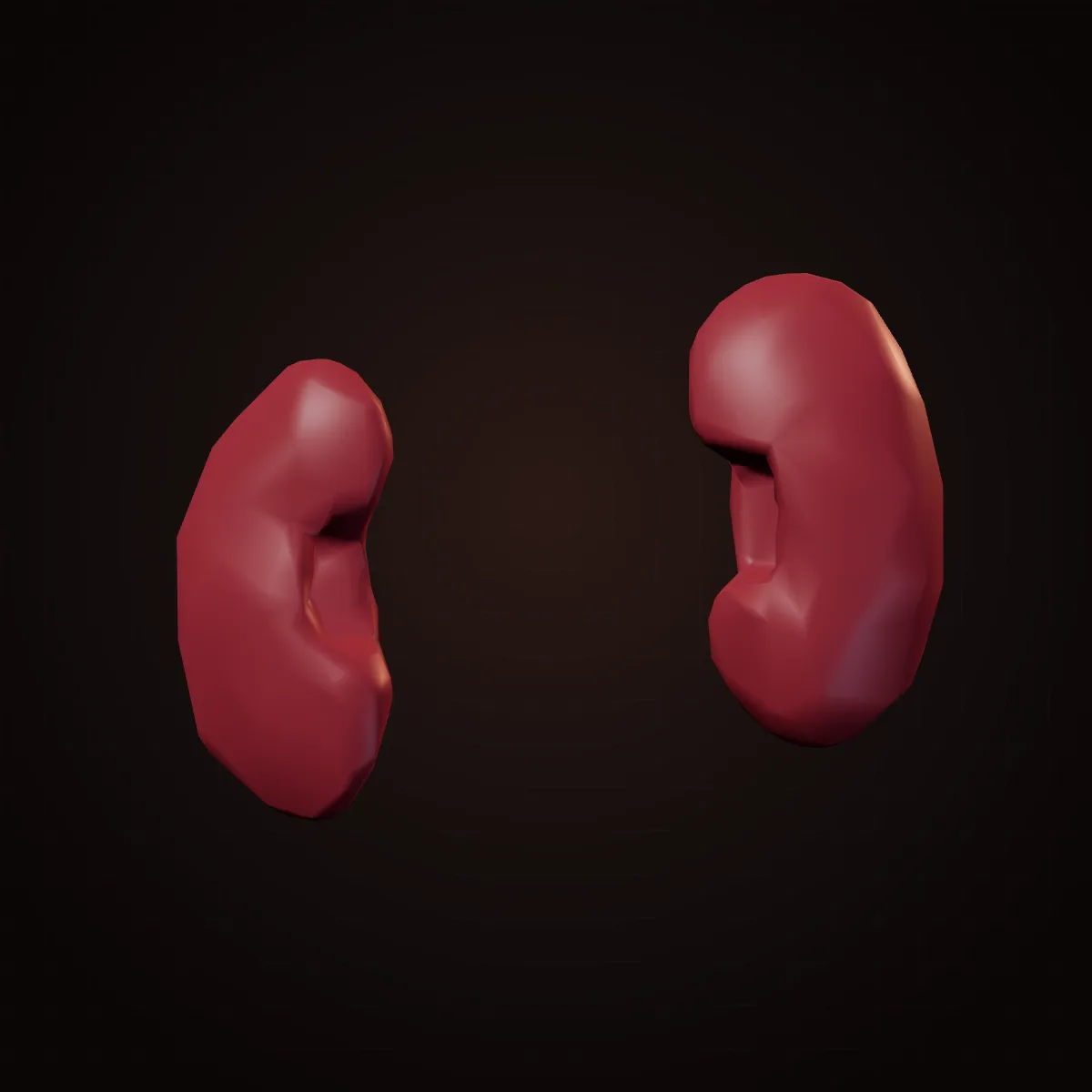
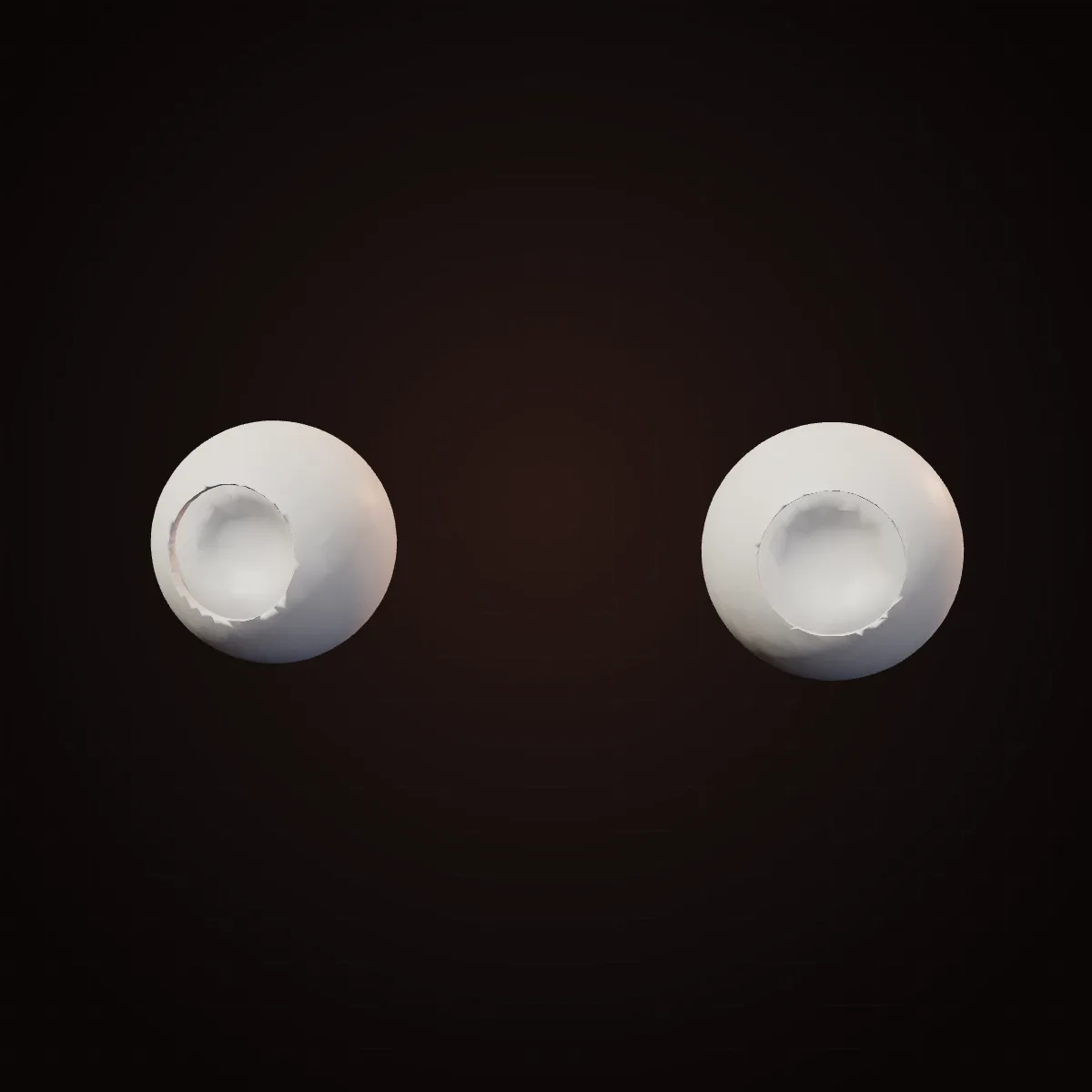
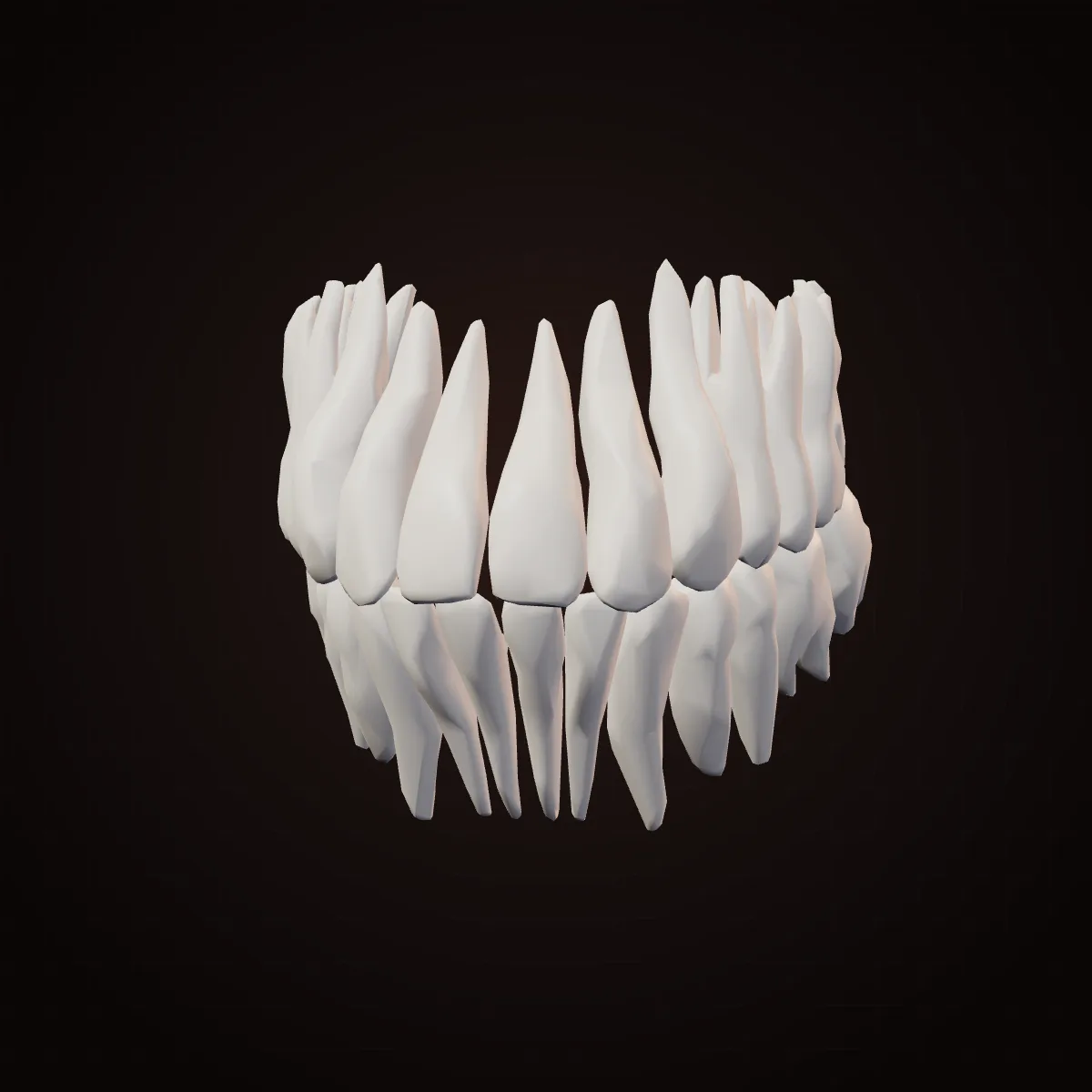
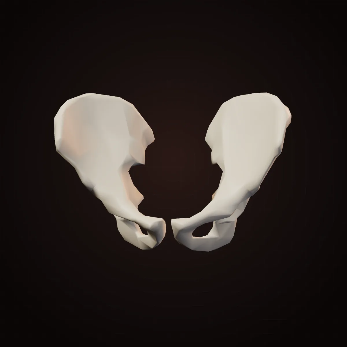
Skeleton31 parts
206 bones in an adult: levers, shields, arches and springs.
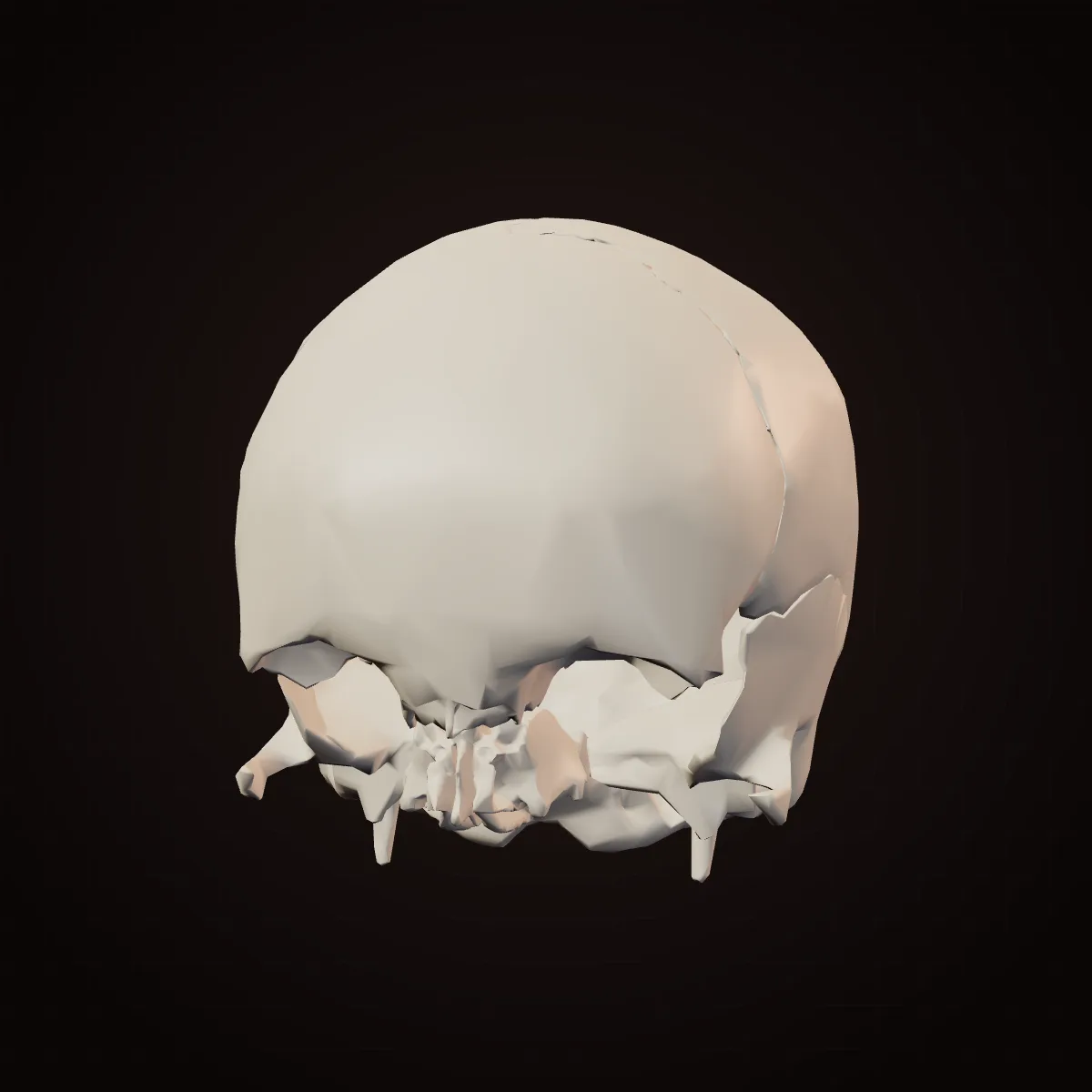
Shape. A rounded dome of curved plates joined by wavy, zig-zag seams called sutures.
Why. A dome encloses the most volume for the least material and spreads a blow across the whole curve instead of concentrating it at one point. The interlocking sutures resist being pulled apart. At birth the plates are not yet fused and are separated by soft spots (fontanelles), so the head can mould through the birth canal and the skull can keep growing with the brain.
Could it be elsewhere? Only partly, or with a trade-off. The vault itself cannot be anywhere else — it is the brain's container. But after an emergency operation to relieve brain swelling, surgeons sometimes store the removed piece of skull inside a pocket of the patient's own belly fat, where it stays alive until it is put back weeks later.
Shape. A frame of thin plates and thick vertical struts around the eye sockets, nose and mouth, hollowed out by air-filled sinuses.
Why. Chewing loads travel up from the teeth through thick pillars of bone (around the nose, the cheekbones and behind the eyes) into the skull, like the buttresses of a building. Between those pillars the bone can be thin and hollow, which keeps the face light. The cone-shaped eye sockets protect the eyes while letting them look forward.
Could it be elsewhere? No — it only works where it is. No. The face is built around the eyes, nose and mouth; the bones are shaped by those openings and by the chewing forces that pass through them.
Shape. A U-shaped (horseshoe) bone with an upright branch at each end that hinges in front of the ear.
Why. It is the only skull bone that moves. The U shape holds the lower teeth in an arch that meets the upper teeth, and the upright branches give the powerful chewing muscles a long lever to pull on. Its joint (the temporomandibular joint) both hinges and slides forward, which is why the mouth can open wide.
Could it be elsewhere? Yes — surgeons already move it. The jaw itself cannot move, but it can be rebuilt from elsewhere. After cancer surgery, surgeons commonly cut a length of the fibula (the thin calf bone), shape it into a jaw and connect its blood supply in the neck.
Shape. A small U-shaped bone in the front of the neck, above the voice box.
Why. It is the only bone in the body that does not form a joint with another bone. It hangs in a sling of muscles and ligaments, giving the tongue and the muscles of swallowing and speech a movable anchor. Being free-floating is the point: it rises and falls every time you swallow.
Could it be elsewhere? No — it only works where it is. No. Its job depends on sitting between the jaw, tongue and voice box.
Shape. Three tiny bones linked in a chain from the eardrum to the inner ear; the stapes, about 3 mm long, is the smallest bone in the body.
Why. Sound in air bounces off fluid, so the inner ear needs a pressure booster. The chain works as a lever, and it funnels vibration from the large eardrum onto the stapes' small footplate. Concentrating the same force on a much smaller area raises the pressure many times over — enough to move the fluid in the cochlea.
Could it be elsewhere? No — it only works where it is. No. They only work between an eardrum and the inner ear's oval window. (Surgeons can replace a stiffened stapes with a tiny prosthesis, but that is a replacement, not a move.)
Shape. A stack of ring-shaped bones with a gentle double S-curve when seen from the side; the vertebrae get bigger towards the bottom.
Why. The curves let the spine act like a spring, absorbing the jolt of each step and keeping the head balanced over the hips. The lower vertebrae carry more of the body's weight, so their bodies are larger. The ring behind each vertebral body forms a protected canal for the spinal cord, and the bony projections are levers for back muscles.
Could it be elsewhere? No — it only works where it is. No. It is the body's central axis; everything else is arranged around it.
Shape. The atlas is a ring with no body; the axis below it has an upward peg (the dens) that sits inside that ring.
Why. The atlas cradles the skull in two cup-shaped surfaces, which allows nodding. The ring turning around the axis' peg works like a pivot, which allows shaking the head. Roughly half of the neck's side-to-side rotation happens at this one joint.
Could it be elsewhere? No — it only works where it is. No. The design is specific to joining the skull to the spine.
Shape. Flat, round cushions between the vertebrae: tough rings of fibre (annulus fibrosus) around a gel-like centre (nucleus pulposus).
Why. The gel centre spreads pressure evenly in all directions, and the layered rings, whose fibres run at alternating angles like a radial tyre, contain it. The result is a shock absorber that also lets the spine bend and twist. When the ring tears, the gel can bulge out and press on a nerve — a 'slipped' disc.
Could it be elsewhere? No — it only works where it is. No. They are shaped to fit between two vertebral bodies.
Shape. The sacrum is a triangular wedge of five fused vertebrae; the coccyx is a small tapering tail below it.
Why. The sacrum is wedged between the two hip bones like the keystone of an arch, passing the weight of the upper body to the pelvis and legs. Fusion makes it rigid. The coccyx is what remains of a tail; in humans it anchors pelvic-floor muscles, ligaments and some fibres of the buttock muscle, and it bears weight when you lean back to sit.
Could it be elsewhere? No — it only works where it is. No. It is load-bearing only as the keystone of the pelvis.
Shape. Curved, flat bones that arc from the spine around the chest and slope downward towards the front.
Why. Curved hoops make a cage that is rigid enough to protect the heart and lungs but flexible enough to absorb a blow. Because they slope down, lifting them swings them up and out — like lifting a bucket handle — which widens the chest and draws air in. The lower 'floating' ribs do not reach the breastbone, leaving the waist free to bend.
Could it be elsewhere? Sometimes — it can develop elsewhere. Sometimes. A small minority of people have an extra 'cervical rib' growing from the lowest neck vertebra, which usually causes no trouble but can press on nerves and vessels to the arm. Surgeons also take rib cartilage to build new ears and noses.
Shape. A flat, dagger-shaped plate: the handle (manubrium), the blade (body) and a small tip (xiphoid process).
Why. It is the front anchor of the rib cage. Flexible strips of cartilage connect it to the ribs, so the chest can expand. Its flat shape shields the heart, and it is close to the skin, which is why it is used for chest compressions and, in adults, for bone-marrow sampling.
Could it be elsewhere? No — it only works where it is. No. Its job is to close the front of the rib cage.
Shape. A long, slender bone with a gentle S-curve lying horizontally between the breastbone and the shoulder.
Why. It is a strut that holds the shoulder out to the side so the arm can swing freely. It is the only bony connection between the arm and the trunk. The S-curve adds springiness; when a fall on an outstretched hand sends force up the arm, the clavicle often breaks first — it is one of the most commonly fractured bones.
Could it be elsewhere? No — it only works where it is. No. It works only as a brace between the sternum and the shoulder blade.
Shape. A thin, triangular plate with a ridge (the spine) and a shallow socket (the glenoid) at its outer corner.
Why. It floats on the back of the rib cage, held by muscles rather than joints, so it can glide and rotate to aim the shoulder socket wherever the arm needs to go. The broad flat surfaces give 17 muscles room to attach. The socket is shallow, which gives the shoulder the widest range of motion of any joint — at the cost of being the joint that dislocates most often.
Could it be elsewhere? No — it only works where it is. No. Its whole design is about steering the shoulder joint.
Shape. A long bone with a smooth ball-shaped head at the top and a pulley-like end at the elbow.
Why. A long bone is a hollow tube with thick walls: bending loads are carried by the outer wall, so a tube is almost as strong as a solid rod but much lighter. The rounded head rotates in the shoulder socket, and the spool-shaped lower end lets the elbow work as a hinge.
Could it be elsewhere? No — it only works where it is. No. Its ends are shaped for the shoulder and elbow joints specifically.
Shape. Two parallel long bones. The ulna is wide at the elbow, the radius is wide at the wrist.
Why. Two bones instead of one allow rotation: the radius can roll over the ulna, turning the palm up (supination) or down (pronation) — the movement of turning a key or a doorknob. The ulna forms a firm hinge at the elbow, and the radius carries the hand at the wrist.
Could it be elsewhere? No — it only works where it is. No. The rotating pair depends on joints at both the elbow and the wrist.
Shape. Eight small, roughly cube-shaped bones in two rows, forming an arch.
Why. Many small bones gliding against each other let the wrist bend and tilt in several directions while still carrying load. The arch they make, bridged by a strong ligament, forms the carpal tunnel, a protected passage for the tendons and the median nerve to the hand. Swelling in that tight tunnel is what causes carpal tunnel syndrome.
Could it be elsewhere? No — it only works where it is. No. Their shapes interlock only with each other, the forearm and the hand.
Shape. Five palm bones fanning out from the wrist, then three short bones in each finger and two in the thumb.
Why. Short segments with hinge joints let fingers curl around objects of any shape. The fan arrangement spreads the fingers for a wide grip. There are no muscles in the fingers themselves: they are moved by long tendons from forearm muscles, which keeps the fingers slim.
Could it be elsewhere? No — it only works where it is. No — but toes can become fingers. See the thumb.
Shape. A short, thick digit set at an angle to the palm, with a saddle-shaped joint at its base.
Why. A saddle joint lets the thumb swing in two planes, so its pad can rotate to meet the pad of each finger (opposition). That one movement is the basis of both a precision pinch and a power grip.
Could it be elsewhere? Yes — surgeons already move it. Yes. When a thumb is lost, surgeons can transplant the patient's own big toe or second toe to the hand, reconnecting bone, tendons, nerves and blood vessels. The toe works as a new thumb.
Shape. A ring-like basin of bone with broad flaring wings at the top and deep cup-shaped sockets on each side.
Why. It transfers the weight of the upper body to the legs, holds the organs of the lower belly, and anchors the big walking muscles. Its broad, curved wings keep the trunk balanced over one leg with each step. In females the inlet is usually wider and rounder and the angle under the pubic bones wider, which leaves room for childbirth.
Could it be elsewhere? No — it only works where it is. No. It is the junction between the spine and the legs. (Surgeons do take bone from the iliac crest for grafts elsewhere.)
Shape. The longest and strongest bone, with a ball-shaped head set on an angled neck, and two rounded knuckles at the knee.
Why. The neck angles the shaft inward so the knees sit under the body's centre of gravity, which makes walking efficient. The ball sits deep in the hip socket for stability. Inside the ends, spongy bone struts line up with the stress lines from standing and walking.
Could it be elsewhere? No — it only works where it is. No. Its shape is built around the hip and knee.
Shape. A flat, rounded triangle of bone embedded in the tendon of the thigh muscle.
Why. It is the largest sesamoid bone — a bone that grows inside a tendon. By holding the tendon away from the knee joint, it gives the thigh muscle a longer lever, so straightening the knee takes less force. It also protects the front of the joint and stops the tendon rubbing on bone.
Could it be elsewhere? No — it only works where it is. No. It only works inside the tendon where it crosses the knee.
Shape. Two C-shaped pads of tough cartilage, thick at the outer edge and thin towards the middle.
Why. The round knuckles of the femur sit on the fairly flat top of the shin bone. The wedge-shaped menisci deepen that surface so the bones fit better, spread the load over a larger area and absorb shock. A twist under load can tear them — one of the most common knee injuries.
Could it be elsewhere? No — it only works where it is. No. They are shaped to the gap between femur and tibia.
Shape. A thick long bone with a flat top (the tibial plateau) and a sharp front edge you can feel under the skin.
Why. It carries almost all the weight passing from the knee to the ankle, so it is thick and roughly triangular in cross-section, which resists bending. Its flat top makes the platform for the knee, and its lower end forms the inner ankle bone.
Could it be elsewhere? No — it only works where it is. No. It is the leg's main weight-bearing column.
Shape. A thin, long bone beside the tibia, ending in the outer ankle bone.
Why. It carries little body weight. Its jobs are to anchor leg muscles and to form the outer wall of the ankle joint, which stops the foot rolling outward.
Could it be elsewhere? Yes — surgeons already move it. Yes. Because most of its shaft is not needed for standing, surgeons routinely remove a length of fibula with its blood vessels and use it to rebuild a jaw or other bones (a 'free fibula flap').
Shape. Seven blocky bones; the heel bone (calcaneus) is the largest, and the talus sits on it like a rocker under the shin.
Why. The talus transfers the body's weight from the leg to the foot and acts as the ankle's hinge; unusually, no muscles attach to it. The big heel bone takes the first impact of every step and gives the Achilles tendon a long lever. Together the tarsals form the back of the foot's arches.
Could it be elsewhere? No — it only works where it is. No. They are shaped for the load path from leg to ground.
Shape. A honeycomb-like lattice of tiny bony struts inside the ends of long bones and inside vertebrae.
Why. The struts line up along the directions of the forces the bone feels, so the bone is strong exactly where it is loaded and light everywhere else — the same idea engineers use in trusses. Bone keeps rebuilding to match its loads (Wolff's law), which is why exercise strengthens bones and space flight weakens them.
Could it be elsewhere? No — it only works where it is. It is found in every bone end already; the pattern is local to each bone's loads.
Shape. Soft tissue filling the spaces inside bones: red marrow (blood-forming) in the spongy bone of the hips, spine, ribs and skull; yellow fatty marrow in long-bone shafts.
Why. Blood-forming stem cells are sensitive, and making billions of blood cells a day needs a protected, well-supplied space. The inside of bone is both. In adults, red marrow is concentrated in the trunk bones.
Could it be elsewhere? Yes — surgeons already move it. Yes, in a striking way. In a bone-marrow (stem cell) transplant, donor cells are simply dripped into a vein. They travel through the blood and settle in the recipient's bones on their own — a process called homing.
Shape. A thin, smooth, glassy layer covering the ends of bones inside joints.
Why. Bathed in joint fluid, it gives one of the lowest-friction surfaces known, and it squashes slightly to spread load. It has no blood vessels, which keeps it smooth but also means it heals very poorly — the root of osteoarthritis.
Could it be elsewhere? Only partly, or with a trade-off. Surgeons can move small plugs of healthy cartilage from a lightly loaded part of a joint to a damaged spot, but only within joints.
Shape. Short bands and cords of dense collagen connecting bone to bone, such as the cruciate ligaments crossing inside the knee.
Why. Collagen fibres are very strong when pulled but bend freely, so ligaments hold joints together while allowing movement in the right directions. The two cruciate ligaments in the knee cross in an X, which stops the shin sliding forward or backward.
Could it be elsewhere? Yes — surgeons already move it. A torn knee ligament (the ACL) is usually rebuilt from a strip of the patient's own tendon taken from the kneecap or hamstrings.
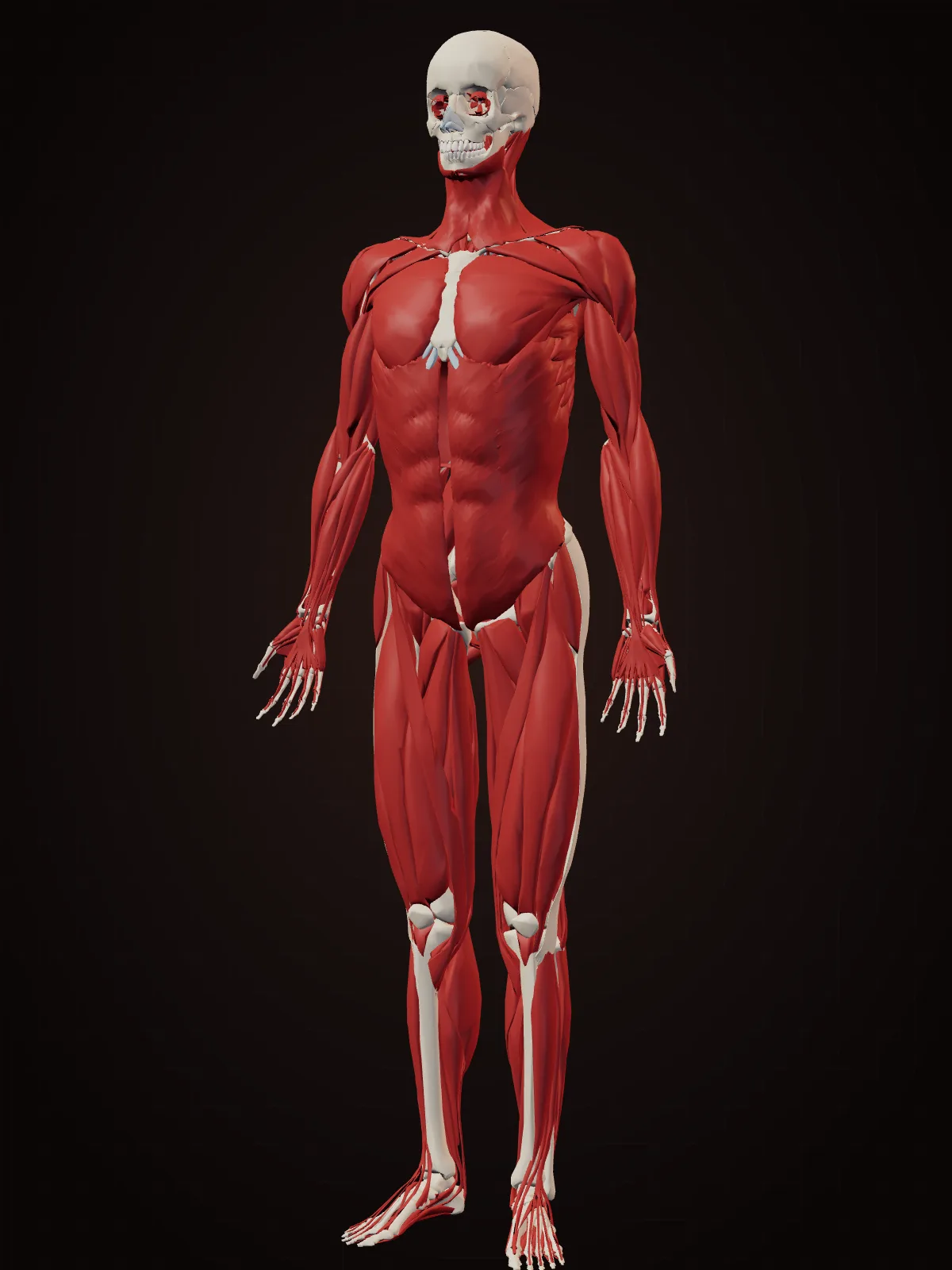
Muscles25 parts
Over 600 skeletal muscles, plus smooth and heart muscle.
Shape. Bundles within bundles: long muscle cells (fibres) packed into fascicles, wrapped in sheets of connective tissue that merge into tendons. Some muscles are spindle-shaped, some strap-like, some feather-like (pennate).
Why. A muscle can only pull, never push, so muscles work in opposing pairs. Fibre arrangement trades speed for force: long parallel fibres shorten a long way quickly, while feather-like fibres set at an angle pack more fibres into the same space and pull harder over a shorter distance.
Could it be elsewhere? Yes — surgeons already move it. Yes, often. Surgeons move whole muscles with their blood supply as 'flaps' — for example, a back muscle to rebuild a breast, or a thigh muscle to a face to restore a smile after nerve paralysis.
Shape. A thick triangular muscle (named after the Greek letter delta, Δ) capping the shoulder.
Why. Its front, middle and back fibres wrap the shoulder from three sides, so it can lift the arm forward, sideways or backward depending on which part contracts. Its rounded bulk also gives the shoulder its shape.
Could it be elsewhere? No — it only works where it is. No. Its job is moving the shoulder joint.
Shape. A large fan-shaped muscle spreading from the collarbone and breastbone and converging on the upper arm.
Why. A fan lets one muscle pull in many directions: the upper fibres lift the arm forward, the lower fibres pull it down and in. The fibres twist as they converge on the humerus, which keeps them working well across the arm's wide range of motion.
Could it be elsewhere? Only partly, or with a trade-off. It can be moved as a flap to cover wounds in the neck and chest, but its pulling job is local.
Shape. A spindle-shaped muscle on the front of the upper arm with two heads (hence 'bi-ceps') joining into one tendon at the elbow.
Why. It bends the elbow, but its tendon attaches to the radius, so it is also the strongest muscle that turns the palm upward. That is why a screwdriver turns more powerfully clockwise for most right-handers.
Could it be elsewhere? No — it only works where it is. No. Its two jobs depend on crossing both the shoulder and the elbow.
Shape. A three-headed muscle on the back of the upper arm.
Why. It is the opposite of the biceps: it straightens the elbow. Three heads give it more mass and one head crosses the shoulder too, so it also helps pull the arm back.
Could it be elsewhere? No — it only works where it is. No.
Shape. Fleshy muscles near the elbow that narrow into long, thin tendons running across the wrist to the fingers.
Why. Putting the bulky muscle in the forearm and sending only slim tendons to the hand keeps the fingers light and nimble — like a puppeteer working strings from a distance. Small muscles inside the hand handle fine adjustments.
Could it be elsewhere? Yes — surgeons already move it. Yes, functionally. In tendon transfer surgery, a working forearm tendon is rerouted to take over a paralysed one.
Shape. A small muscle with a long, thin tendon down the middle of the inner wrist; you can see it by touching thumb to little finger and bending the wrist.
Why. It tightens the tissue of the palm, a job more important in animals that use their forelimbs to walk or climb. In humans it is so minor that grip strength is about the same without it.
Could it be elsewhere? Yes — surgeons already move it. Yes — it is a favourite spare part. Surgeons take its tendon to repair or replace tendons and ligaments elsewhere, for example in the hand or elbow.
Shape. Two long vertical straps down the front of the belly, divided across by bands of tendon into blocks.
Why. It bends the trunk forward and holds the belly wall firm. The tendon bands split each strap into short segments, which helps the muscle hold its position and resist bulging. The blocks are visible only when the fat layer over them is thin.
Could it be elsewhere? Yes — surgeons already move it. Yes, partly. A strip of it with its blood supply is a classic flap for rebuilding a breast or filling wounds in the pelvis.
Shape. Three thin, broad sheets of muscle wrapping the side of the belly, with fibres running in different directions in each layer.
Why. Like plywood, layers running at different angles make a thin wall strong in every direction. Together they hold the organs in, twist the trunk and squeeze the belly for coughing, pushing and breathing out hard.
Could it be elsewhere? No — it only works where it is. No. They are the belly wall.
Shape. A large, flat, kite-shaped muscle from the base of the skull and the spine out to each shoulder blade.
Why. Its upper, middle and lower fibres pull the shoulder blade up, in and down, steering it under the arm. It also supports the weight of the arms, which is one reason it aches with stress and long hours at a desk.
Could it be elsewhere? No — it only works where it is. No.
Shape. The broadest back muscle, a wide triangular sheet from the lower back that twists into a narrow tendon on the upper arm.
Why. Its wide origin and narrow insertion give it a long pulling line to draw the arm down and back — the movement of a pull-up, rowing or swimming.
Could it be elsewhere? Yes — surgeons already move it. Yes. It is one of the most used muscle flaps in surgery, moved with its blood supply to rebuild a breast or cover large wounds; the body compensates well for its loss.
Shape. A thin, dome-shaped sheet of muscle separating the chest from the belly, with a tendon in the middle.
Why. When the dome contracts it flattens and moves down, enlarging the chest so air flows into the lungs. Relaxing lets it spring back up. The dome shape is what turns a simple contraction into a big change of volume. Openings let the oesophagus, the aorta and the main vein pass through.
Could it be elsewhere? No — it only works where it is. No. It works only as the floor of the chest.
Shape. A thick, broad, square-ish muscle forming the shape of the buttock.
Why. It straightens the hip — climbing stairs, standing up, sprinting — and keeps the trunk from pitching forward when you run. It is much larger in humans than in other apes, which researchers link to upright walking and long-distance running.
Could it be elsewhere? Only partly, or with a trade-off. Parts of it are used as flaps to close pressure sores, but its job is local to the hip.
Shape. Four muscles on the front of the thigh that join into one tendon around the kneecap.
Why. Four muscles sharing one tendon pool their pull to straighten the knee, which holds you up with each step and launches every jump. The kneecap in their tendon increases their leverage.
Could it be elsewhere? No — it only works where it is. No.
Shape. Three long muscles on the back of the thigh, from the sit bone to below the knee.
Why. Crossing both hip and knee, they straighten the hip and bend the knee, and they brake the swinging leg at the end of each running stride — the moment when they are most often torn.
Could it be elsewhere? Yes — surgeons already move it. Two of their tendons are a standard graft for rebuilding a torn knee ligament (ACL).
Shape. A long, thin strap running diagonally across the front of the thigh, from hip to inner knee.
Why. It is the longest muscle in the body. Its diagonal path lets it bend and rotate both hip and knee at once — the cross-legged sitting position. Its name comes from the Latin for tailor.
Could it be elsewhere? No — it only works where it is. No.
Shape. Bulging calf muscles that taper into the Achilles tendon, the thickest and strongest tendon in the body, attached to the heel.
Why. The calf lifts the heel to push you forward. The long tendon stretches as you land and recoils as you push off, returning stored energy like a spring, which makes walking and running cheaper. A tendon in the leg and a heavy muscle near the knee also keeps the lower leg light to swing.
Could it be elsewhere? No — it only works where it is. No.
Shape. A thick, rectangular muscle on the side of the jaw that you can feel bulge when you clench your teeth.
Why. It runs from the cheekbone to the angle of the jaw, close to the back teeth, so it acts with short leverage on the molars — producing very high biting force where grinding happens. It is often described as one of the strongest muscles for its size.
Could it be elsewhere? No — it only works where it is. No.
Shape. Thin sheets and rings of muscle just under the skin; the eyes and mouth are each circled by a ring muscle (orbicularis).
Why. Unlike most skeletal muscles, they attach to skin rather than only to bone, so they move the skin itself — that is what lets a face make expressions. The ring muscles close the eyes and lips like drawstrings.
Could it be elsewhere? No — it only works where it is. No. They are tied to the skin of the face.
Shape. The smallest skeletal muscle in the body, a few millimetres long, attached to the stapes in the middle ear.
Why. When a sound is very loud, it contracts and stiffens the ossicle chain, damping the vibration reaching the inner ear. That reflex gives some protection against sustained loud noise, though it is too slow to protect against sudden blasts.
Could it be elsewhere? No — it only works where it is. No.
Shape. A strap muscle running diagonally up the side of the neck from the breastbone and collarbone to behind the ear.
Why. Working alone, it turns the head to the opposite side; working together, the pair bend the neck. Its diagonal line also makes it a landmark that divides the neck into front and back zones for doctors.
Could it be elsewhere? No — it only works where it is. No.
Shape. A hammock- or bowl-shaped sheet of muscle across the bottom of the pelvis, with openings for the urethra, vagina (in females) and anus.
Why. Standing upright puts the weight of the belly organs on the bottom of the pelvis. A muscular hammock holds them up while staying able to relax for passing urine and stool, and stretching hugely in childbirth.
Could it be elsewhere? No — it only works where it is. No. It is the floor of the trunk.
Shape. Spindle-shaped cells with no stripes, arranged in sheets in the walls of the gut, blood vessels, airways, bladder and uterus.
Why. Wrapped in rings and lengthways layers around tubes, it squeezes slowly and tirelessly without conscious control — pushing food along in waves (peristalsis) and tightening or widening blood vessels.
Could it be elsewhere? No — it only works where it is. It is already everywhere a tube needs to squeeze.
Shape. Rings of muscle around a passage — at the stomach's exit, the bladder outlet, the anus, the iris of the eye.
Why. A ring that tightens closes a tube completely and evenly from all sides, like a drawstring, and opens when it relaxes. The body uses this one design to control flow wherever it needs a valve made of muscle.
Could it be elsewhere? No — it only works where it is. Each one is placed where a passage needs controlling.
Shape. Sheets of connective tissue that wrap muscles, group them into compartments and connect them to bone and skin.
Why. It keeps muscles in place, lets them slide past each other and transmits their pull. Tight compartments of fascia are why swelling after a leg injury can become dangerous: the pressure has nowhere to go.
Could it be elsewhere? Yes — surgeons already move it. Strips of fascia from the thigh are used as grafts to repair hernias, eyelids and other tissues.
Nervous system15 parts
Brain, spinal cord and the nerves that wire them to everything.
Shape. Two wrinkled hemispheres whose surface is a thin sheet of grey matter (the cortex) folded into ridges (gyri) and grooves (sulci).
Why. Thinking happens in the cortex, so more surface means more processing. Folding packs a large sheet into a skull small enough to be born through the pelvis — roughly two-thirds of the cortex is hidden inside the folds. Folding also keeps connected areas close together, shortening wiring.
Could it be elsewhere? No — it only works where it is. No. It needs the skull's protection, a huge blood supply and short paths to the eyes, ears and spinal cord.
Shape. A fist-sized structure at the back and bottom of the brain with very fine, tightly packed parallel folds (folia).
Why. It fine-tunes movement, balance and timing by comparing what you meant to do with what your body actually did. That calculation needs enormous numbers of small neurons arranged in a repeating circuit, and the very fine folding fits them in.
Could it be elsewhere? No — it only works where it is. No. It sits beside the brainstem where signals from the body and from the cortex meet.
Shape. A stalk joining the brain to the spinal cord.
Why. Every signal between brain and body passes through this narrow stalk, and it runs breathing, heart rate, swallowing and wakefulness automatically. Ten of the twelve pairs of cranial nerves connect to it. Its compact central position is why even a small injury here can be life-threatening.
Could it be elsewhere? No — it only works where it is. No.
Shape. A thick, arched band of nerve fibres joining the left and right hemispheres.
Why. With hundreds of millions of fibres, it lets the two halves share information so you experience one world. Its arched shape follows the curve of the hemispheres above the brain's fluid spaces.
Could it be elsewhere? No — it only works where it is. No.
Shape. A curved, rolled-up ridge deep in each temporal lobe; its name is Greek for 'seahorse', which it resembles.
Why. It is a fold of cortex rolled in on itself, which places its layers of neurons in a loop that is well suited to linking pieces of an experience into a memory and mapping places. It is essential for forming new long-term memories.
Could it be elsewhere? No — it only works where it is. No.
Shape. An almond-shaped cluster of neurons in front of each hippocampus (amygdala is Greek for almond).
Why. It tags experiences with emotional weight, especially threat. Its position next to the memory system and with fast links to the senses lets it trigger a fear response before you are consciously aware of the danger.
Could it be elsewhere? No — it only works where it is. No.
Shape. Three membranes wrap the brain and spinal cord — tough dura mater, web-like arachnoid, delicate pia mater — with clear fluid between the inner two.
Why. The brain is soft. Floating in fluid makes it buoyant: a brain of about 1.4 kg presses down with the weight of only about 50 g, so it does not crush its own lower parts or its blood vessels. The fluid also cushions knocks and carries away waste.
Could it be elsewhere? No — it only works where it is. No.
Shape. A cable about as thick as a finger running inside the spine, ending around the first or second lumbar vertebra in adults; below that a bundle of nerve roots continues like a horse's tail (cauda equina).
Why. It is the trunk line between brain and body and handles fast reflexes by itself. It is shorter than the spine because the spine keeps growing after the cord stops, which is why doctors can take a sample of spinal fluid with a needle low in the back without hitting the cord.
Could it be elsewhere? No — it only works where it is. No. It needs the spine's bony tunnel.
Shape. Cells with a bushy tree of input branches (dendrites) and one long output cable (axon) ending in many small terminals.
Why. A branching tree lets one neuron collect signals from thousands of others; the axon carries its single answer, sometimes a very long way. The motor neurons that move your toes run from the lower spine all the way to the foot — among the longest cells in the body.
Could it be elsewhere? No — it only works where it is. They are already everywhere. Most neurons cannot divide, which is why nerve damage heals so slowly.
Shape. Layers of fatty membrane wound around an axon like tape, with tiny gaps (nodes of Ranvier) at intervals.
Why. Insulation stops the electrical signal leaking, and the signal jumps from gap to gap instead of crawling along. That makes conduction many times faster — up to about 100 m/s in the fastest fibres. Diseases that strip myelin, such as multiple sclerosis, slow or block signals.
Could it be elsewhere? No — it only works where it is. It wraps axons wherever speed matters.
Shape. Cables of many axons grouped in bundles and wrapped in connective tissue, branching out to every part of the body.
Why. Like an electrical cable, bundling protects many separate signal wires and lets them travel together before branching off to their targets. The layered wrapping gives nerves the strength to stretch a little as joints move.
Could it be elsewhere? Yes — surgeons already move it. Yes. Surgeons repair gaps in important nerves with a graft from a less important one — usually the sural nerve, which carries skin sensation from the outer foot.
Shape. The largest and thickest nerve in the body, running from the lower spine through the buttock and down the back of the thigh.
Why. It carries almost all signals to and from the leg and foot, so it bundles a huge number of fibres. Its route under the buttock muscle explains why a pinched root in the lower back can send pain all the way down the leg (sciatica).
Could it be elsewhere? No — it only works where it is. No.
Shape. A long nerve running from the brainstem down the neck and into the chest and belly, branching to the throat, heart, lungs and gut ('vagus' is Latin for wandering).
Why. It is the main line of the 'rest and digest' system, slowing the heart and stimulating digestion, and most of its fibres carry information from the organs back to the brain. One long wandering nerve can reach every organ along the way.
Could it be elsewhere? No — it only works where it is. No.
Shape. A branch of the vagus that goes down into the chest, hooks under a major artery (the aorta on the left), and climbs back up to the voice box.
Why. It is a famous example of history shaping anatomy. In fish ancestors the nerve ran straight past a gill artery. As the neck lengthened and the heart moved down into the chest, the nerve stayed looped under the artery, so it had to grow longer. It works, but its route is a detour, not an optimal design.
Could it be elsewhere? Sometimes — it can develop elsewhere. Sometimes. In a small number of people with an unusual right-side artery, the right nerve does not loop down at all and goes straight to the larynx — a 'non-recurrent' nerve that surgeons check for during thyroid surgery.
Shape. A nerve that runs through a groove behind the inner elbow, just under the skin.
Why. To get past the elbow without being stretched each time the arm bends, it passes behind the joint's pivot point. That route leaves it with little padding, so knocking it gives the electric tingle down to the little finger.
Could it be elsewhere? Yes — surgeons already move it. Surgeons can move it to the front of the elbow (an ulnar nerve transposition) when it keeps getting compressed.
Senses19 parts
Eyes, ears, nose and tongue: instruments shaped by physics.
Shape. A nearly spherical ball about 24 mm across, kept firm by fluid pressure inside.
Why. A sphere can rotate in its socket in any direction while the optics stay aligned. Fluid pressure holds the precise distance between the lens and the retina; if the eye is slightly too long or short, the image is out of focus — the cause of short- and long-sightedness.
Could it be elsewhere? No — it only works where it is. No. It needs the bony socket, the eye muscles and a short optic nerve path to the brain.
Shape. A clear, curved dome at the front of the eye, with no blood vessels.
Why. The bend of light at its curved surface does about two-thirds of the eye's focusing. It has no blood vessels so it can stay perfectly clear; it gets oxygen straight from the air and from the fluid behind it. Laser eye surgery works by reshaping this curve.
Could it be elsewhere? Yes — surgeons already move it. A related surprise: for some people blinded by severe corneal scarring, surgeons build a new window for the eye from the patient's own tooth and jawbone holding a plastic lens — the osteo-odonto-keratoprosthesis, or 'tooth-in-eye' operation.
Shape. A clear, flexible, convex disc behind the iris, held by fine fibres.
Why. A curved, convex lens bends light to a focus. Being elastic, it changes shape: rounder for near objects, flatter for far ones. With age it stiffens, so most people need reading glasses in their forties. When it clouds (a cataract), surgeons replace it with a plastic lens.
Could it be elsewhere? No — it only works where it is. No.
Shape. A coloured ring of muscle around a round opening.
Why. It works like a camera's aperture: a ring muscle shrinks the pupil in bright light and radial muscles widen it in the dark. A smaller opening also sharpens focus, which is why you squint to see clearly.
Could it be elsewhere? No — it only works where it is. No.
Shape. A thin sheet of light-sensitive cells lining the back of the eye, with a small pit (the fovea) in the middle and a spot with no receptors where the optic nerve leaves.
Why. Lining the curved back of the eyeball keeps the whole image in focus. In vertebrates the wiring lies in front of the light-sensing cells, so the nerve fibres must pass through the retina to leave the eye, creating a blind spot; the brain fills it in. The receptors face the back because they depend on the blood-rich layer behind them.
Could it be elsewhere? No — it only works where it is. No. Octopus eyes, which evolved separately, have their wiring behind the receptors and no blind spot — proof that the arrangement is history, not necessity.
Shape. A tiny pit in the centre of the retina packed only with cone cells.
Why. Other layers of the retina are pushed aside, so light reaches the receptors without passing through anything. This is where you see sharpest and in colour, which is why your eyes move constantly to point the fovea at whatever you are looking at.
Could it be elsewhere? No — it only works where it is. No.
Shape. Folds of thin skin stiffened by a plate of dense tissue, edged with curved lashes.
Why. Blinking wipes tears across the cornea to keep it clean and wet. Lashes trigger a blink when touched and alter airflow over the eye; a 2015 study across mammals found lashes are typically about one-third of the eye's width, a length that reduces evaporation and dust reaching the surface.
Could it be elsewhere? No — it only works where it is. No.
Shape. Arched strips of short hair on the brow ridge, growing outward.
Why. The arch and the sideways direction of the hairs channel sweat and rain to the sides of the face, away from the eyes. They are also highly mobile and important in expressions and in recognising faces.
Could it be elsewhere? Yes — surgeons already move it. Hair from the back of the scalp can be transplanted to rebuild eyebrows.
Shape. An almond-sized gland above the outer corner of each eye, and tiny drainage holes at the inner corners leading to a duct into the nose.
Why. Tears flow from the outer corner across the eye and drain at the inner corner, so blinking sweeps the surface in one direction. The drain empties into the nose — which is why your nose runs when you cry.
Could it be elsewhere? No — it only works where it is. No.
Shape. A curved, folded flap of elastic cartilage and skin around the ear canal.
Why. It funnels sound in, and its ridges and hollows echo sound slightly differently depending on whether it comes from above, below, in front or behind. The brain reads those tiny differences to tell where a sound is coming from, especially its height.
Could it be elsewhere? Yes — surgeons already move it. Yes, during reconstruction. In 2018 US Army surgeons shaped rib cartilage into an ear, grew it under the skin of a soldier's forearm to give it blood vessels, then attached it to her head.
Shape. An S-shaped tube about 2.5 cm long, closed at the inner end by a thin cone-shaped membrane (the tympanic membrane).
Why. A tube closed at one end resonates. The ear canal's length boosts sounds around 2–5 kHz, the range that carries many speech sounds. The curve protects the eardrum from direct hits, and wax and tiny hairs trap dust. The cone-shaped drum vibrates as one piece and drives the ossicles.
Could it be elsewhere? No — it only works where it is. No.
Shape. A fluid-filled, snail-shaped tube coiled about two and a half turns, about the size of a pea.
Why. Inside, a membrane runs the length of the tube; it is stiff and narrow at the base, so high pitches vibrate it there, and floppy and wide at the tip, where low pitches do. Coiling packs a tube of about 3.5 cm into a tiny space in the hardest bone of the skull. A cochlear implant works by placing electrodes along this pitch map.
Could it be elsewhere? No — it only works where it is. No.
Shape. Three fluid-filled loops set roughly at right angles to each other, like the corner of a box.
Why. Three perpendicular loops cover the three directions of rotation — nodding, tilting and turning. When the head turns, fluid lags behind and bends sensory hairs. Two small sacs nearby sense gravity and straight-line acceleration. A mismatch between these signals and vision causes motion sickness.
Could it be elsewhere? No — it only works where it is. No.
Shape. A narrow tube from the middle ear to the back of the nose, usually closed.
Why. It opens when you swallow or yawn to equalise air pressure on both sides of the eardrum — the 'pop' on a plane. In young children it is shorter and more horizontal, so germs travel up it more easily, which is one reason ear infections are common in childhood.
Could it be elsewhere? No — it only works where it is. No.
Shape. A projecting, pyramid-shaped structure of bone and cartilage with two downward-facing nostrils.
Why. Downward nostrils and a projecting nose slow and direct the air into the nasal cavity. Researchers have found that nostril width tends to vary with the temperature and humidity of the climate a population's ancestors lived in — an adaptation for warming and moistening air.
Could it be elsewhere? Yes — surgeons already move it. Yes, during reconstruction. A classic technique swings a flap of forehead skin down to make a new nose; surgeons have also shaped a nose and grown it on a patient's forehead or arm before moving it into place.
Shape. Scroll-shaped shelves of thin bone covered with moist, blood-rich lining, projecting into each side of the nasal cavity.
Why. Scrolls make the air swirl over a large, warm, wet surface, so by the time it reaches the lungs it is near body temperature, humidified and filtered. Their linings swell and shrink in turn, so one nostril usually carries more air than the other for a few hours (the nasal cycle).
Could it be elsewhere? No — it only works where it is. No.
Shape. A small patch of lining in the roof of each side of the nose, with nerve endings passing up through a sieve-like bone into the brain.
Why. Placing it high and out of the main airflow protects it, and sniffing swirls odours up to it. Its nerve cells reach straight through the skull, the shortest route to the brain of any sense. Humans have roughly 400 kinds of working smell receptor.
Could it be elsewhere? No — it only works where it is. No.
Shape. Air-filled hollows in the bones around the nose — in the forehead, cheeks and between and behind the eyes — lined with mucous membrane.
Why. Their exact purpose is still debated. Proposed roles include making the skull lighter, adding resonance to the voice, adding a crumple zone for the face and helping humidify air. What is clear is that they drain into the nose through small openings, which block easily with a cold.
Could it be elsewhere? No — it only works where it is. No.
Shape. The tongue is covered in small bumps (papillae); taste buds sit in the walls of many of them.
Why. The bumps grip and move food, and grooves around some of them hold dissolved food against the taste cells. The old 'tongue map' — sweet at the tip, bitter at the back — is a myth: all five basic tastes can be detected across the tongue.
Could it be elsewhere? No — it only works where it is. No — though some taste buds also sit on the palate and throat.
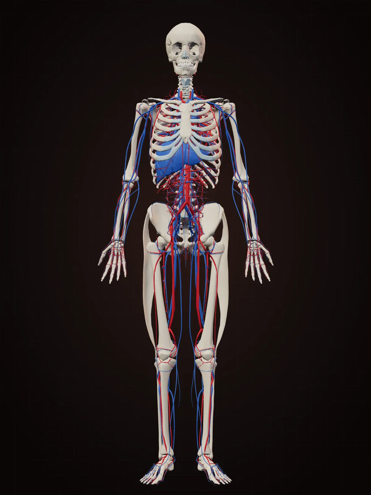
Heart & blood11 parts
A pump, a pipe network and the cells that ride in it.
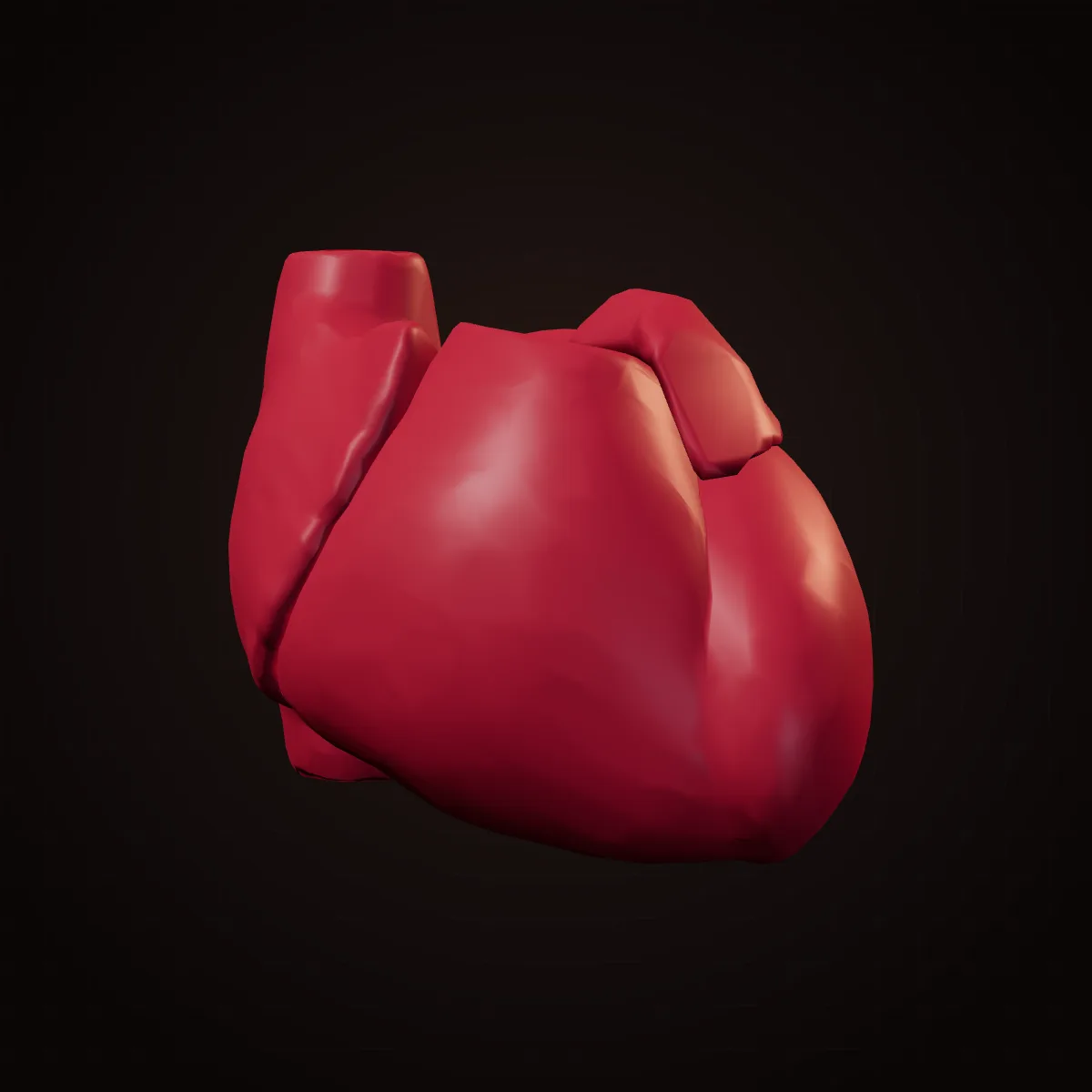
Shape. A muscular cone about the size of a fist, lying behind the breastbone with its pointed tip (apex) angled down and to the left.
Why. It is two pumps side by side: the right sends blood a short way through the lungs, the left sends it around the whole body, so the left ventricle's wall is much thicker. The muscle fibres wrap in spirals, so each beat wrings the heart like a towel rather than just squeezing it — emptying it more completely. Thin-walled upper chambers (atria) collect blood; thick lower ones (ventricles) push it out.
Could it be elsewhere? Only partly, or with a trade-off. Remarkably, yes, in a limited way. In a 'piggyback' (heterotopic) heart transplant, first done in Cape Town in 1974, a donor heart is placed in the right side of the chest alongside the patient's own heart and connected to help it pump. It is rarely done today, but it worked. In a small number of people the heart also develops on the right side (dextrocardia).
Shape. Thin flaps (cusps) at the exits of each chamber; the two between the upper and lower chambers are tethered by cord-like tendons to small muscles.
Why. They let blood flow only one way. The cusps are shaped like pockets or parachutes: backflow fills them and pushes them shut. The tendon cords stop the flaps from blowing inside-out when the ventricles squeeze hard. The 'lub-dub' heartbeat is the sound of the valves closing.
Could it be elsewhere? No — it only works where it is. No. Replacement valves (mechanical, or from pig or cow tissue) are put in the same place.
Shape. Arteries that leave the base of the aorta and branch over the surface of the heart like a crown (corona).
Why. The heart muscle cannot use the blood passing through its chambers; it needs its own supply. Branching over the outer surface lets them feed the wall from outside in. A blockage in one starves the muscle beyond it — a heart attack.
Could it be elsewhere? Yes — surgeons already move it. In a bypass operation, surgeons take a vein from the leg or an artery from inside the chest wall or the forearm and use it to route blood around a blockage.
Shape. A tough, double-layered sac around the heart with a thin film of fluid between the layers.
Why. The fluid lets the heart beat inside it with almost no friction, and the tough outer layer anchors the heart in place and stops it overfilling.
Could it be elsewhere? No — it only works where it is. No.
Shape. The largest artery, about as wide as a garden hose, rising from the heart, arching over and running down in front of the spine.
Why. Its walls are full of elastic fibres. With each beat they stretch, then recoil between beats, smoothing the heart's spurts into steadier flow. The arch turns the blood downward while sending branches up to the head and arms.
Could it be elsewhere? No — it only works where it is. No.
Shape. Thick-walled, round tubes with layers of muscle and elastic tissue, branching into ever smaller vessels.
Why. They carry blood away from the heart at high pressure, so their walls must be strong. Branching means every cell in the body is close to a supply. Muscle in the walls of small arteries controls where blood goes, widening to feed working muscles and narrowing elsewhere.
Could it be elsewhere? Yes — surgeons already move it. Arteries such as the radial artery in the forearm are used as bypass grafts for the heart.
Shape. Thinner, floppier tubes than arteries, with one-way valves inside, especially in the legs.
Why. Blood in veins is at low pressure, so it needs help to get back to the heart against gravity. Leg muscles squeeze the veins as you walk, and the valves stop blood flowing back down. When the valves fail, veins bulge — varicose veins. Veins also act as a reservoir, holding most of the body's blood at rest.
Could it be elsewhere? Yes — surgeons already move it. Yes. The great saphenous vein, running up the inside of the leg, is the most common graft used to bypass blocked heart arteries; the leg's other veins take over its job.
Shape. The smallest vessels, a mesh of tubes about 5–10 micrometres wide with walls one cell thick.
Why. Oxygen, nutrients and wastes can only pass through a very thin wall over a short distance. Capillaries are so narrow that red blood cells pass in single file and touch the wall, and so many that almost every cell is within a short distance of one.
Could it be elsewhere? No — it only works where it is. They are everywhere already; new ones grow wherever tissue needs them.
Shape. Flexible discs, thinner in the middle than at the rim (biconcave), about 7–8 micrometres across, with no nucleus.
Why. The dimpled disc gives a lot of surface for oxygen to pass through and means no part of the cell is far from the surface. Without a nucleus there is more room for haemoglobin. The shape is also very flexible, so the cells fold to squeeze through capillaries narrower than themselves. In sickle cell disease, cells become rigid and curved and get stuck.
Could it be elsewhere? Yes — surgeons already move it. They travel everywhere already; donated red cells work in another person's body after a transfusion.
Shape. White cells are round when floating but change shape to crawl; platelets are small discs that turn spiky when activated.
Why. White blood cells must leave the blood to reach infections, so they flatten and squeeze between the cells of vessel walls. Platelets travel smooth so they do not snag, then sprout projections at a wound so they can stick to each other and plug it.
Could it be elsewhere? No — it only works where it is. They move around the whole body by design.
Shape. The body looks symmetrical outside but not inside: the heart points left, the liver is on the right, the stomach and spleen on the left, and the gut is coiled one consistent way.
Why. In the early embryo, beating tiny hairs (cilia) create a leftward flow of fluid that switches on different genes on each side. That sets the asymmetry for every organ. Asymmetry lets organs of different shapes pack tightly together.
Could it be elsewhere? Sometimes — it can develop elsewhere. Yes — the whole arrangement can be mirrored. In situs inversus totalis, about 1 in 10,000 people, every organ is a mirror image of the usual layout. Many people only find out through a scan for something else.
Breathing7 parts
A branching tree that ends in hundreds of millions of air sacs.
Shape. Two cone-shaped spongy organs filling the chest. The right lung has three lobes; the left has two and a notch where the heart sits.
Why. The lungs are an enormous folded surface packed into the chest. The left is smaller because the heart takes up space on that side. Their soft, elastic tissue stretches when the chest expands and recoils to push air out. Dividing them into lobes means disease or surgery can affect one part without losing the whole lung.
Could it be elsewhere? Only partly, or with a trade-off. They cannot work outside the chest. But a lobe can be moved between people: in a living-donor lobar transplant, two donors each give one lower lobe to a recipient.
Shape. A tube about 10–12 cm long held open by C-shaped rings of cartilage, open at the back.
Why. The rings keep the airway from collapsing when you breathe in hard. They are open at the back, where the oesophagus lies, so a swallowed mouthful can bulge into that space on its way down. The lining is covered in tiny beating hairs that sweep mucus and dust up towards the throat.
Could it be elsewhere? No — it only works where it is. No.
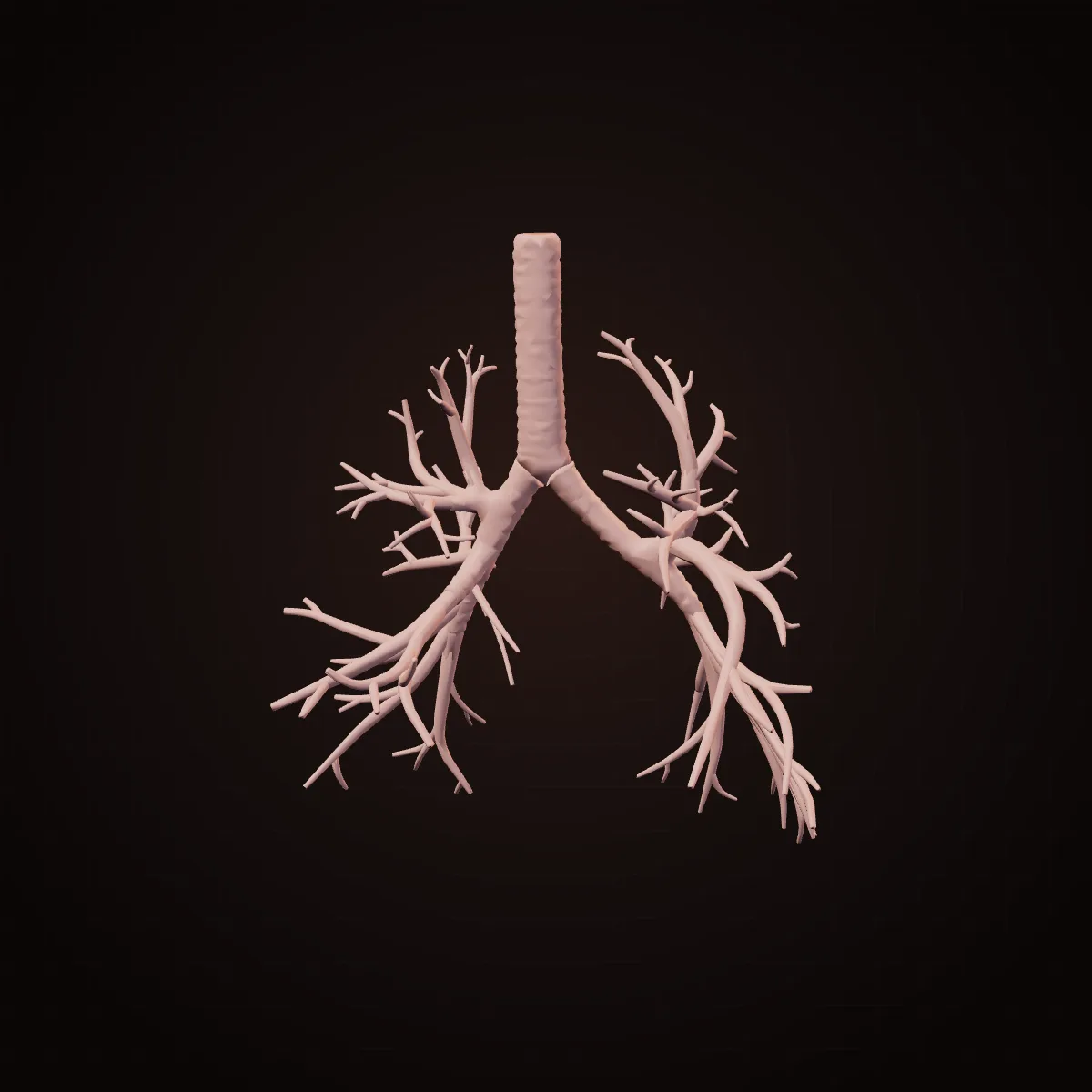
Shape. The windpipe splits into two, and each branch keeps splitting, about 23 times, into ever narrower tubes — an upside-down tree.
Why. Repeated branching is the simplest way to deliver air from one pipe to hundreds of millions of tiny sacs all through the lung. The right main bronchus is wider and more vertical, which is why an inhaled peanut more often lands in the right lung.
Could it be elsewhere? No — it only works where it is. No.
Shape. Tiny bubble-like sacs clustered like grapes at the ends of the smallest airways, each wrapped in capillaries.
Why. Gas exchange needs a huge surface and a very thin barrier. Hundreds of millions of tiny sacs give an area of tens of square metres, and the wall between air and blood is a fraction of a micrometre thick. A slippery coating (surfactant) stops the smallest sacs collapsing; babies born very early may lack it.
Could it be elsewhere? No — it only works where it is. No.
Shape. A box of cartilages at the top of the windpipe; the shield-shaped thyroid cartilage makes the Adam's apple. Inside, two folds of tissue form a V.
Why. Its first job is to protect the airway — the folds slam shut when you swallow or cough. Air pushed through the closed folds makes them vibrate, and that is the voice; tightening them raises the pitch. Testosterone at puberty enlarges the cartilage and lengthens the folds, which lowers the voice and makes the Adam's apple more prominent.
Could it be elsewhere? No — it only works where it is. No.
Shape. A leaf-shaped flap of elastic cartilage behind the tongue.
Why. Air and food share the throat. When you swallow, the larynx rises and the epiglottis tips back over its opening like a lid, steering food into the oesophagus. If food goes 'down the wrong way', a cough clears it.
Could it be elsewhere? No — it only works where it is. No.
Shape. A thin double membrane: one layer covers each lung, the other lines the chest wall, with a film of fluid between.
Why. The fluid lets the lungs slide smoothly, and its surface tension makes the lung stick to the chest wall the way two wet sheets of glass stick together. So when the chest expands, the lung has to follow. If air gets between the layers, the lung collapses (a pneumothorax).
Could it be elsewhere? No — it only works where it is. No.
Digestion16 parts
A tube from mouth to anus, with glands that feed it.
Shape. Incisors are chisels, canines are cones, premolars and molars are broad with bumps (cusps). Each has a crown of enamel, the hardest substance in the body, over softer dentine.
Why. Different shapes for different jobs: incisors cut, canines grip and tear, molars crush and grind. Enamel is mostly mineral, which makes it extremely hard but unable to repair itself, so it must last a lifetime.
Could it be elsewhere? Yes — surgeons already move it. Yes, astonishingly. In the 'tooth-in-eye' operation (osteo-odonto-keratoprosthesis), a patient's canine tooth with a sliver of jawbone is shaped into a mount for a plastic lens, grown under the skin of the cheek for months, then placed into the eye to restore sight.
Shape. The last molars at the back of each side of each jaw, usually appearing in the late teens or twenties.
Why. Human jaws have become shorter over our evolution, and modern soft diets may leave them shorter still, so there is often not enough room for these last teeth. They then come in at an angle or stay stuck in the bone (impacted).
Could it be elsewhere? Sometimes — it can develop elsewhere. Variation is common: roughly one in five people is missing at least one wisdom tooth because it never forms. Dentists can also transplant a healthy wisdom tooth into the gap left by a lost molar.
Shape. A muscular organ with no bone inside, anchored to the jaw, hyoid and skull.
Why. It is a 'muscular hydrostat', like an elephant's trunk or an octopus arm: muscle fibres run lengthways, across and up-down, and because muscle tissue does not compress, squeezing one way makes it extend another. That gives it the shape-changing control needed to move food, swallow and form speech sounds.
Could it be elsewhere? No — it only works where it is. No.
Shape. Clusters of tiny sacs like bunches of grapes, draining through branching ducts into the mouth.
Why. Saliva wets food so it can be swallowed, starts digesting starch, protects teeth and helps you taste. Each gland's ducts empty near the teeth and tongue where the saliva is needed.
Could it be elsewhere? Yes — surgeons already move it. Yes. For people with severe dry eye, surgeons have moved a submandibular gland to the temple and led its duct to the eye, so saliva keeps the eye moist.
Shape. The roof of the mouth: a hard bony front part and a soft muscular back part ending in a small hanging tag (the uvula).
Why. The hard palate gives the tongue a firm surface to press food against and lets babies suckle while breathing. When you swallow, the soft palate lifts to seal off the nose, so food and drink do not come out of it.
Could it be elsewhere? No — it only works where it is. No.
Shape. A muscular tube about 25 cm long, flat when empty, running from the throat behind the windpipe and through the diaphragm to the stomach.
Why. Food is pushed down by rings of muscle squeezing in waves (peristalsis), not by gravity — astronauts can swallow in orbit, and you can swallow upside down. A ring of muscle at the bottom stops stomach acid coming up; when it leaks, you feel heartburn.
Could it be elsewhere? Yes — surgeons already move it. It can be replaced from below. After the oesophagus is removed for cancer, surgeons often pull the stomach up into the chest and join it to the throat, or use a piece of colon as a new food pipe.
Shape. A J-shaped muscular bag under the left ribs, with deep folds (rugae) lining it when empty.
Why. It is a storage tank and mixer: the folds flatten out so it can stretch to hold a large meal, three layers of muscle running in different directions churn food with acid and enzymes, and a ring of muscle at the exit releases it a little at a time. A thick layer of mucus protects the wall from its own acid.
Could it be elsewhere? Yes — surgeons already move it. Yes, partly. It can be pulled up into the chest to replace a removed oesophagus, and people can live without a stomach altogether by eating small, frequent meals.
Shape. A narrow tube several metres long, coiled in the middle of the belly, its lining folded into ridges covered with tiny finger-like villi.
Why. This is where most nutrients are absorbed, and absorption needs surface. Folds, then villi, then microscopic microvilli on each cell multiply the area. A 2014 study estimated the inner surface of the whole gut at about 32 square metres, mostly in the small intestine — around half a badminton court, not the 'tennis court' of older textbooks.
Could it be elsewhere? Yes — surgeons already move it. Pieces of it are routinely moved. Surgeons can make a new bladder from a length of small intestine, or use it to rebuild the oesophagus or vagina.
Shape. Villi are finger-like projections about a millimetre long; each lining cell carries a brush of thousands of microscopic microvilli.
Why. Each villus contains capillaries and a lymph vessel right under a single layer of cells, so nutrients only travel a tiny distance into the blood. Coeliac disease flattens the villi, which is why it causes poor absorption.
Could it be elsewhere? No — it only works where it is. No.
Shape. A wider tube that frames the small intestine — up the right side, across, and down the left — puckered into pouches (haustra).
Why. It absorbs water and salts and houses most of the body's gut bacteria. The pouches slow the contents and give bacteria time to ferment fibre. Running around the edge of the belly lets it be fixed to the back wall while the small intestine stays mobile in the middle.
Could it be elsewhere? Yes — surgeons already move it. A segment of colon can be moved up into the chest to replace the oesophagus.
Shape. A narrow, blind-ended tube a few centimetres long hanging from the start of the large intestine, low on the right.
Why. It contains a lot of immune tissue, and one widely discussed hypothesis is that it acts as a safe house for good gut bacteria to repopulate the colon after illness. Its narrow, dead-end shape is also why it can block and become inflamed (appendicitis). People live normally without it.
Could it be elsewhere? Yes — surgeons already move it. Yes. In the Mitrofanoff procedure, surgeons reconnect the appendix between the bladder and the belly button, turning it into a channel through which a person can empty their bladder with a catheter.
Shape. The final stretch of bowel, which can expand to store stool, ending in a short canal closed by two rings of muscle.
Why. The inner ring works automatically; the outer ring is under your control. Two rings let the body sense that the rectum is filling and still choose when to empty it. Cushions of blood vessels help seal the canal, and their swelling causes piles (haemorrhoids).
Could it be elsewhere? Only partly, or with a trade-off. After some operations the bowel can be brought out through the belly wall as a stoma, which works but without a sphincter's control.
Shape. A large wedge, thick on the right and tapering to the left, tucked under the diaphragm and protected by the lower ribs.
Why. It is the body's chemical processing plant. Blood from the intestines goes to the liver first through the portal vein, so nutrients and toxins are handled before they reach the rest of the body. Its wedge shape fills the dome under the right side of the diaphragm. It is the largest internal organ.
Could it be elsewhere? Yes — surgeons already move it. Partly. A living donor can give part of their liver, and both parts regrow to near full size within months. In rare 'auxiliary' transplants, a partial donor liver is placed alongside the patient's own.
Shape. A small pear-shaped sac tucked under the liver on the right.
Why. The liver makes bile all the time, but you only need a lot of it after a fatty meal. The gallbladder stores and concentrates bile, then squeezes it into the intestine when food arrives. Concentrated bile can form stones, which is why the gallbladder is so often removed — and people manage fine without it.
Could it be elsewhere? No — it only works where it is. No — but it is optional; bile then flows straight from the liver.
Shape. A long, soft, tadpole-shaped gland lying across the back of the belly behind the stomach, its head tucked into the C-shaped curve of the duodenum.
Why. It does two jobs. Most of it makes digestive juice, poured into the duodenum exactly where food leaves the stomach. Scattered through it are about a million clusters of hormone cells (islets) that make insulin and glucagon. Its deep position protects it.
Could it be elsewhere? Yes — surgeons already move it. Yes. Transplanted pancreases are usually placed low in the belly and connected to the vessels of the pelvis, not where the original sits. Islets alone can be infused into the liver's portal vein, where they lodge and make insulin.
Shape. The peritoneum is a slippery membrane lining the belly; the mesentery is a fan-shaped double fold of it that holds the intestines to the back wall.
Why. The fan lets several metres of intestine attach along a short root at the back of the belly, while carrying their blood vessels, lymph vessels and nerves. The slippery surface lets loops of bowel slide over each other as they move. In 2016 researchers proposed classifying the mesentery as a single continuous organ.
Could it be elsewhere? No — it only works where it is. No.
Reproductive10 parts
Organs built around temperature, transport and protection.
Shape. Two oval organs, each about 4–5 cm long, packed with coiled tubes, hanging outside the body in the scrotum.
Why. Sperm production works best a few degrees below core body temperature — roughly 2–4 °C cooler — so the testes sit outside the trunk. Each is packed with hundreds of metres of tightly coiled tubes where sperm form, and a net of veins around the artery cools incoming blood before it arrives.
Could it be elsewhere? Sometimes — it can develop elsewhere. They start inside the belly near the kidneys and normally descend before birth. In a few percent of full-term baby boys one has not finished descending (undescended testis); most come down in the first months, and the rest are brought down by surgery, partly because a testis left inside the body produces sperm poorly.
Shape. A pouch of thin, wrinkled skin with a layer of muscle, divided inside into two compartments.
Why. It is a thermostat. In the cold, its muscle wrinkles the skin and a muscle around each cord pulls the testes up towards the body for warmth; in the heat, they relax and the testes hang lower. Thin skin with many sweat glands loses heat well.
Could it be elsewhere? No — it only works where it is. No.
Shape. Three cylinders of spongy erectile tissue — two on top, one underneath carrying the urethra — wrapped in tough sheaths and skin.
Why. When the spaces in the spongy tissue fill with blood, the tough outer sheath stops them expanding, so pressure rises and the organ becomes rigid — a hydraulic design. The lower cylinder stays softer so the urethra inside it stays open for semen to pass.
Could it be elsewhere? Yes — surgeons already move it. It can be rebuilt from a flap of forearm or thigh tissue (phalloplasty), and a small number of penis transplants have been performed.
Shape. A walnut-sized gland directly below the bladder, wrapped around the urethra.
Why. It adds fluid to semen, and its muscle helps push semen out. Because it encircles the urethra, when it enlarges with age — which is very common — it squeezes the tube and makes urinating slower and more frequent.
Could it be elsewhere? No — it only works where it is. No.
Shape. Two almond-shaped organs, each about 3–5 cm long, on either side of the uterus.
Why. They hold a lifetime supply of immature eggs, each in a tiny fluid-filled sac (follicle), and make oestrogen and progesterone. Each month a few follicles grow and usually one releases its egg from the surface towards the waiting fallopian tube.
Could it be elsewhere? Yes — surgeons already move it. Partly, yes. Ovarian tissue frozen before cancer treatment has been grafted back not only to the pelvis but also to sites such as the forearm or belly wall, where it has restarted hormone production.
Shape. Narrow tubes from the uterus towards each ovary, ending in a trumpet-shaped opening fringed with finger-like projections (fimbriae).
Why. The tube is not attached to the ovary. Instead the fringes sweep over it, and beating hairs and muscle waves draw the released egg in and carry it towards the uterus. Fertilisation usually happens in the tube.
Could it be elsewhere? Sometimes — it can develop elsewhere. This shows why location matters: in an ectopic pregnancy, an embryo implants outside the uterus, usually in the tube. A tube cannot stretch like a uterus, so it is a medical emergency.
Shape. A hollow, upside-down pear about 7–8 cm long, with a thick muscular wall and a narrow neck (the cervix) opening into the vagina.
Why. Its lining rebuilds every month to receive an embryo. The thick muscle wall grows enormously in pregnancy and then contracts with great force in labour. The narrow, mucus-plugged cervix keeps the inside protected and then widens for birth.
Could it be elsewhere? No — it only works where it is. No. Uterus transplants (the first resulting birth was in Sweden in 2014) place the donor uterus in the same position, connected to the pelvic vessels.
Shape. A stretchy muscular canal with folded walls from the cervix to the outside; the external structures include the labia and the clitoris.
Why. Folds (rugae) let it stretch greatly during childbirth. Its naturally acidic environment, kept by resident bacteria, helps protect against infection. The clitoris is built from erectile tissue like the penis; both develop from the same embryonic structure.
Could it be elsewhere? No — it only works where it is. No — though it can be reconstructed using skin grafts or a segment of bowel.
Shape. Glands made of lobes, each a cluster of milk-producing sacs like a bunch of grapes, drained by branching ducts to the nipple, set in fat.
Why. A branching duct system lets thousands of milk sacs empty through a handful of openings at the nipple. Most of the breast's size is fat, not gland, which is why size does not predict milk supply.
Could it be elsewhere? Sometimes — it can develop elsewhere. Sometimes. In the embryo, breast tissue can form anywhere along a 'milk line' from the armpit to the groin. A few percent of people have an extra nipple or small patch of breast tissue somewhere along it, most often just under the breast.
Shape. A temporary, flat, disc-shaped organ about 20–25 cm across at birth, attached to the inside of the uterus, with the umbilical cord at its centre.
Why. It is a huge surface of tree-like branches soaked in the mother's blood, so oxygen and nutrients pass to the baby's blood without the two bloods mixing. It also makes pregnancy hormones. It is the only organ the body builds, uses for nine months and then discards.
Could it be elsewhere? No — it only works where it is. It attaches where the embryo implants. When it lies too low and covers the cervix (placenta praevia), delivery has to be by caesarean.
Hormone glands8 parts
Small glands that signal through the blood.
Shape. A pea-sized gland hanging on a stalk under the brain, sitting in a saddle-shaped hollow of bone (the sella turcica, 'Turkish saddle').
Why. Often called the master gland, it controls the thyroid, adrenals, ovaries and testes and makes growth hormone. Its stalk links it directly to the hypothalamus, so the brain can turn hormone signals up or down quickly. The bony saddle protects it.
Could it be elsewhere? No — it only works where it is. No. It must be connected to the hypothalamus.
Shape. An almond-sized region at the base of the brain, just above the pituitary.
Why. It is where the nervous system and hormones meet. It senses temperature, salt and blood sugar and controls hunger, thirst, sleep and body temperature, partly by commanding the pituitary directly below it.
Could it be elsewhere? No — it only works where it is. No.
Shape. A tiny pine-cone-shaped gland deep in the middle of the brain.
Why. It releases melatonin at night, signalling darkness to the body. It receives its information about light indirectly, through a chain of nerves from the eyes — which is why bright screens at night can delay sleep.
Could it be elsewhere? No — it only works where it is. No.
Shape. A butterfly-shaped gland with two lobes joined by a bridge, wrapped around the front of the windpipe below the Adam's apple.
Why. It sets the body's metabolic pace with hormones made using iodine. Its two lobes hug the airway and it moves up when you swallow, which is how doctors examine it. Iodine shortage makes it swell (goitre) — the reason for iodised salt.
Could it be elsewhere? Sometimes — it can develop elsewhere. Sometimes. In the embryo, the thyroid starts at the back of the tongue and descends to the neck. Rarely it stays behind as a 'lingual thyroid' at the base of the tongue, or leaves pieces along the way.
Shape. Four tiny glands, each about the size of a grain of rice, usually on the back of the thyroid.
Why. They control the level of calcium in the blood, which nerves and muscles need to work. Being tiny and separate, they can be overlooked — and their position varies a lot from person to person.
Could it be elsewhere? Yes — surgeons already move it. Yes. When they have to be removed during neck surgery, surgeons can cut a gland into small pieces and implant them into a forearm muscle, where they grow a blood supply and keep making hormone. The forearm is chosen because blood can be sampled from it easily.
Shape. Small glands sitting like caps on top of each kidney: the right roughly triangular, the left more crescent-shaped.
Why. Each is two glands in one. The outer layer (cortex) makes cortisol, aldosterone and some sex hormones; the inner core (medulla) is modified nerve tissue that releases adrenaline in seconds when the nervous system signals danger. Their position next to the kidney is close to the kidney's blood flow, which aldosterone helps regulate.
Could it be elsewhere? No — it only works where it is. No.
Shape. A soft, two-lobed gland behind the breastbone, large in babies and children and shrinking after puberty, mostly replaced by fat in adults.
Why. It is the school where immune T cells mature and learn not to attack the body's own tissues. Most of that training happens early in life, so the organ shrinks once its main work is done.
Could it be elsewhere? No — it only works where it is. No.
Shape. Microscopic clusters of hormone cells scattered through the pancreas like islands, making up only a small fraction of its mass.
Why. Scattering them through a gland with a rich blood supply means insulin and glucagon reach the blood quickly — and go first to the liver, the main organ they control.
Could it be elsewhere? Yes — surgeons already move it. Yes. In islet transplantation, donor islets are infused into the portal vein and settle in the liver, where they make insulin.
Skin, hair & nails12 parts
The largest organ and its keratin tools.
Shape. A layered sheet: a thin outer epidermis of flattened cells that constantly flake off and are replaced, over a thicker dermis of collagen and elastic fibres, nerves and vessels.
Why. The outer layer is a waterproof barrier of dead cells filled with keratin; the dermis gives strength and stretch. The skin is thinnest on the eyelids, where movement matters, and thickest on the palms and soles, which take wear. It is the body's largest organ.
Could it be elsewhere? Yes — surgeons already move it. Yes — the most moved tissue in surgery. Skin grafts are taken from one place, often the thigh, and applied to burns or wounds elsewhere; thin grafts' donor sites regrow in a couple of weeks.
Shape. Fine ridges of skin in loops, whorls and arches on fingertips, palms and soles.
Why. They form in the womb, and the pattern is unique to each person, even identical twins. Their purpose is still being studied: ridges help with grip on rough surfaces and channel away moisture, and they may make the touch sensors under the skin more sensitive to fine textures.
Could it be elsewhere? No — it only works where it is. No. Ridged skin only grows on the palms, soles and digits.
Shape. Shafts of dead keratin growing from follicles in the skin. Straight hair has a round cross-section; curly hair is flatter and grows from a curved follicle.
Why. Scalp hair shields the head from sun and helps keep heat; eyelashes, eyebrows and nose hairs guard openings; body hair improves the sense of touch. Humans have about as many follicles as other apes, but most produce fine, short hairs, which helps us lose heat by sweating.
Could it be elsewhere? Yes — surgeons already move it. Yes. Hair transplants move follicles from the back of the head, which resists male-pattern baldness, to the bald top, and the moved hairs mostly keep their resistance (the 'donor dominance' idea described in 1959).
Shape. Tiny bands of smooth muscle attaching each hair follicle to the skin.
Why. In cold or fear they contract, making hairs stand up and the skin bumpy. In furry mammals that traps warm air or makes the animal look bigger. In humans, with little body hair, the effect is mostly a leftover — though recent research in mice suggests these muscles also help support hair-follicle stem cells.
Could it be elsewhere? No — it only works where it is. No.
Shape. Flat, slightly curved plates of hard keratin on the backs of the fingertips and toes, growing out from a root (the matrix) under the skin fold.
Why. A nail is a rigid backing for the soft fingertip: when you press on something, the pad pushes against the nail, which improves precise touch and grip and helps pick up small objects. Nails protect the fingertips and are handy tools for scratching and peeling. They replaced claws in our primate ancestors.
Could it be elsewhere? Only partly, or with a trade-off. Not as a whole, but nail-bed tissue taken from a toe can be grafted to rebuild a damaged fingernail bed.
Shape. Coiled tubes deep in the skin with a long duct to a pore. Eccrine glands are everywhere; apocrine glands are in the armpits and groin.
Why. Evaporating sweat carries heat away. Humans have unusually many eccrine glands, and together with our sparse hair this makes us among the best-cooled mammals — a key to our ability to keep moving in the heat. Apocrine glands produce a richer sweat that skin bacteria turn into body odour.
Could it be elsewhere? No — it only works where it is. No.
Shape. Small sac-like glands opening into hair follicles, most numerous on the face and scalp, absent from palms and soles.
Why. They release an oily substance (sebum) that waterproofs and softens skin and hair. Blocked follicles with excess sebum are the start of acne, which is why it appears where these glands are most dense.
Could it be elsewhere? No — it only works where it is. No.
Shape. Clusters of large round cells each filled with a single droplet of fat, under the skin and around organs.
Why. Fat stores energy compactly, insulates, and pads areas that take pressure, such as the heel and the palm. It is also a hormone-making organ (leptin tells the brain how much energy is stored). Fat around the organs behaves differently and is more linked to disease than fat under the skin.
Could it be elsewhere? Yes — surgeons already move it. Yes. Fat grafting moves fat, by liposuction and injection, from the belly or thighs to fill out the face, breast or scars.
Shape. Cells with long branches at the base of the epidermis, passing packets of pigment (melanin) to the skin cells around them.
Why. The branches let one melanocyte supply pigment to dozens of neighbouring cells, and the pigment forms a cap over each cell's nucleus, shielding its DNA from ultraviolet light. Skin colour varies mainly with how much and what kind of melanin is made, not with the number of melanocytes.
Could it be elsewhere? Yes — surgeons already move it. In vitiligo, surgeons can transplant pigment cells from normal skin to white patches.
Shape. Soft folds around the mouth built on a ring muscle, with a red border where the skin is very thin.
Why. They seal the mouth for eating, drinking and sucking, and shape speech sounds. The red colour comes from blood vessels showing through very thin skin; lips have no sweat glands or hair and few oil glands, which is why they dry and chap easily. They are also packed with touch receptors.
Could it be elsewhere? No — it only works where it is. No.
Shape. A scar in the middle of the belly, folded in or out, where the umbilical cord was attached.
Why. Before birth, the cord carried the baby's blood to and from the placenta. After it is cut, the stump dries and falls off, and the healed scar is the navel. Whether it is an 'innie' or 'outie' depends on how the scar heals.
Could it be elsewhere? No — it only works where it is. No — but surgeons sometimes recreate one in a new position during belly reconstruction.
Shape. Small features that are reduced in humans: a pink fold at the inner corner of the eye, muscles that let some people wiggle their ears, a small bump on the rim of some ears.
Why. They match fully working structures in other animals — a third eyelid in many mammals and birds, muscles that turn the ears towards sounds in cats and dogs. Reduced does not always mean useless: the fold in the eye still helps drain tears. They are evidence of our evolutionary history.
Could it be elsewhere? No — it only works where it is. No. They only make sense in the place they came from.