 By
Quick answer: Why is every part of the human body shaped the way it is?
- Protect — domes, cages and fluid cushions: skull, ribs, cerebrospinal fluid.
- Transport — tubes with pumps and one-way valves: heart, veins, gut, ureters.
- Exchange — folds and branching trees that pack huge surfaces into small spaces: lungs, gut, kidneys.
- Move and carry — levers, arches and springs: long bones, the foot, the Achilles tendon.
- Sense — precision instruments shaped by physics: the cochlea's spiral, the eye's sphere, the ear's lever chain.
Key takeaways
- About 18 shapes explain almost every part of the body; the same answer (a tube, a fold, an arch) appears in very different organs.
- Surface area is the body's most expensive design goal: alveoli and gut folds exist to multiply it.
- Location is part of the design. A kidney works low in the belly after a transplant; a cochlea works nowhere but inside the skull.
- Surgeons already move many parts: toes become thumbs, the fibula becomes a jaw, a tooth becomes a mount for a lens in the eye.
- Evolution builds on what came before, so some designs are detours, not optimal: the eye's blind spot and the looping laryngeal nerve.
Pick up a pen and think about what your hand just did. Twenty-seven bones moved, the thumb swung across the palm on a saddle-shaped joint, and the force came from muscles in your forearm pulling on tendons as thin as string, so your fingers could stay slim. None of those shapes is decoration. Each is the answer to a physical problem, and the body uses the same small set of answers again and again. This guide explains that design logic part by part. Alongside it, we built an interactive 3D human body explorer on a real anatomical scan — BodyParts3D, 2,211 structures reconstructed from one adult male14 — where you can rotate the body, peel back its layers and click any of 164 parts to see its shape, why that shape works, and whether it could sit anywhere else.
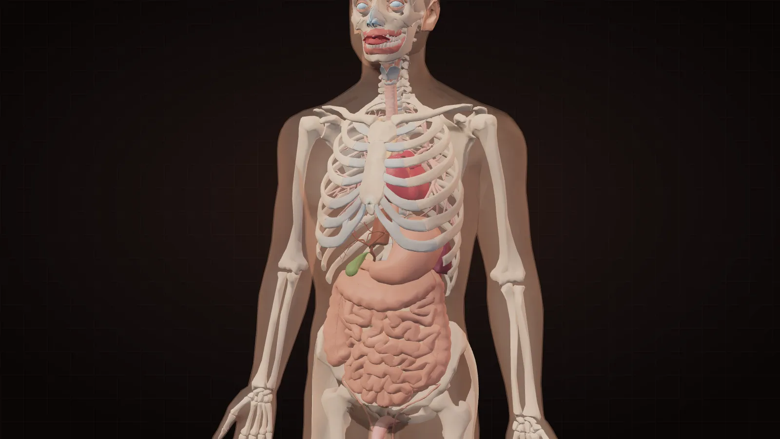

Why is every body part shaped the way it is?
Every body part is shaped to solve a physical problem — protecting, transporting, exchanging, carrying load or sensing — and only a few geometries solve each problem well. That is why a skull and a diaphragm are both domes, why blood vessels, airways and the gut are all tubes, and why lungs, kidneys and intestines are folded or branched.
Biologists put it as "form follows function", and anatomy textbooks organise the whole body this way: bone shapes are classified as long, short, flat, irregular and sesamoid precisely because each shape does a different job1. The deeper reason is physics. A dome spreads a blow. A tube with a pump moves fluid. Diffusion only works over tiny distances, so any organ that exchanges gases or nutrients needs an enormous surface packed into a small space — and the only ways to do that are folding and branching.
When we sorted all 164 parts in the explorer by their dominant geometry, the same patterns kept returning:
| Design pattern | What it solves | Parts in the explorer | Examples |
|---|---|---|---|
| Cable | Carry pull or signals over distance | 22 | Tendons, nerves, hair, spinal cord |
| Plate | Shield organs, anchor muscles | 16 | Scapula, sternum, skin, nails |
| Tube | Move something along one path | 14 | Oesophagus, aorta, veins, ureters |
| Sac | Store, then empty | 13 | Bladder, stomach, gallbladder, lungs |
| Ring | Open, close or hold a passage | 12 | Sphincters, iris, windpipe rings |
| Lever | Turn small pulls into big movements | 11 | Femur, humerus, jaw, finger bones |
| Branching tree | Reach every point from one trunk | 9 | Bronchi, arteries, neurons |
| Spiral | Coil something long into a small space | 9 | Cochlea, myelin, outer ear |
| Cone | Funnel force, sound or flow to a point | 9 | Heart, teeth, ear canal |
| Folded surface | Pack huge area into small space | 7 | Cortex, small intestine, alveoli |
| Arch | Carry load with little material | 7 | Foot arches, ribs, wrist bones |

Table: The most common design patterns among the 164 parts in the Science of Apps human body explorer (one dominant pattern per part)
Six shapes from the 3D scan
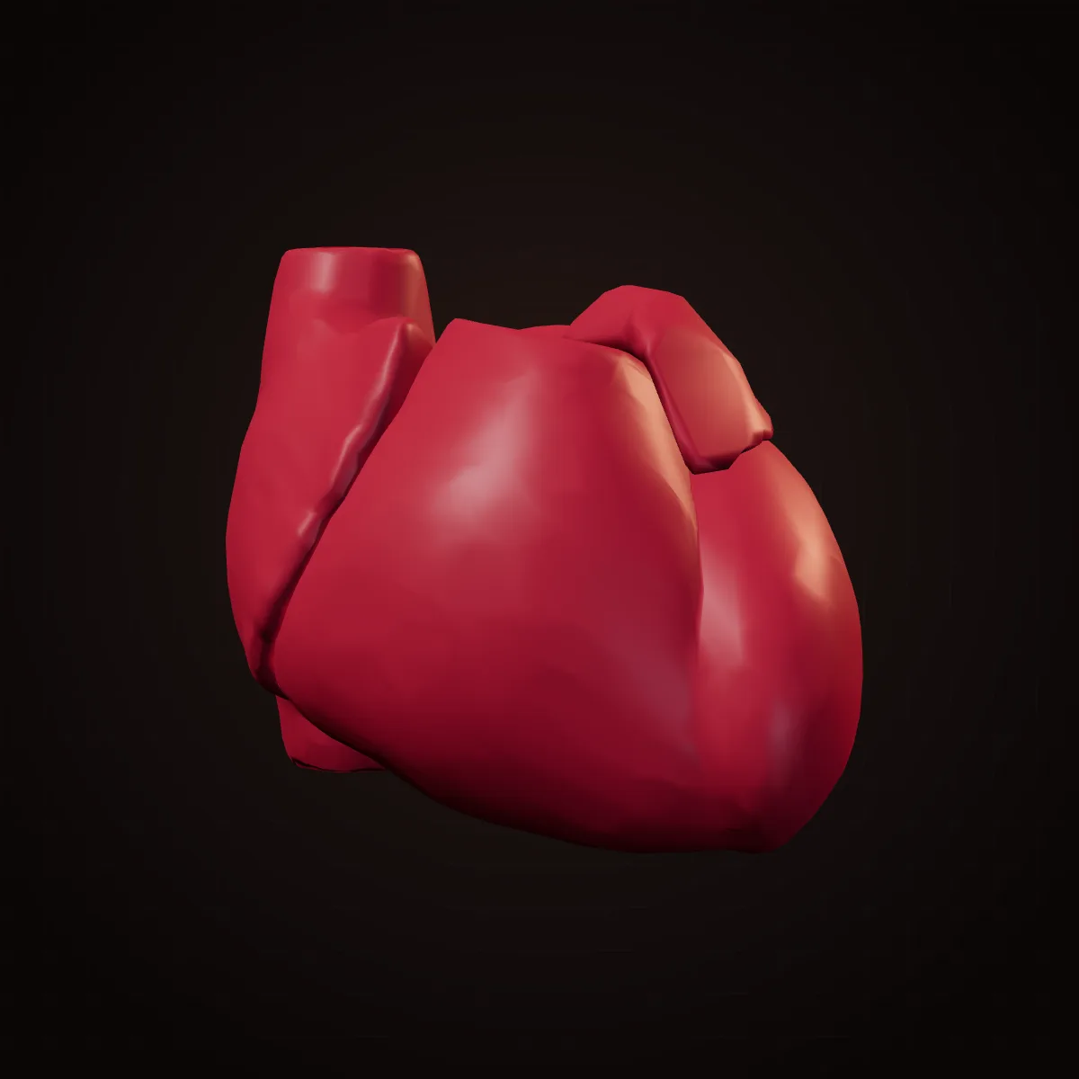

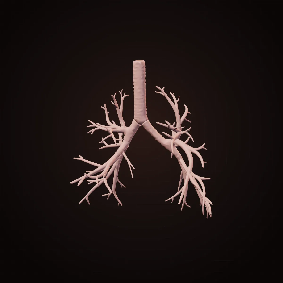
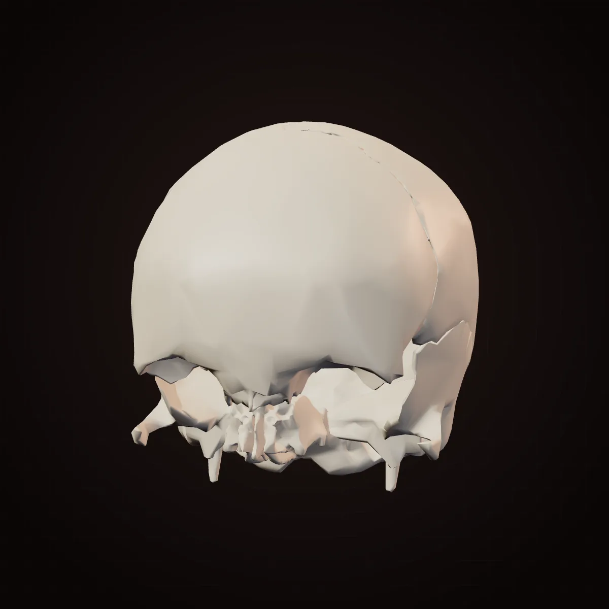
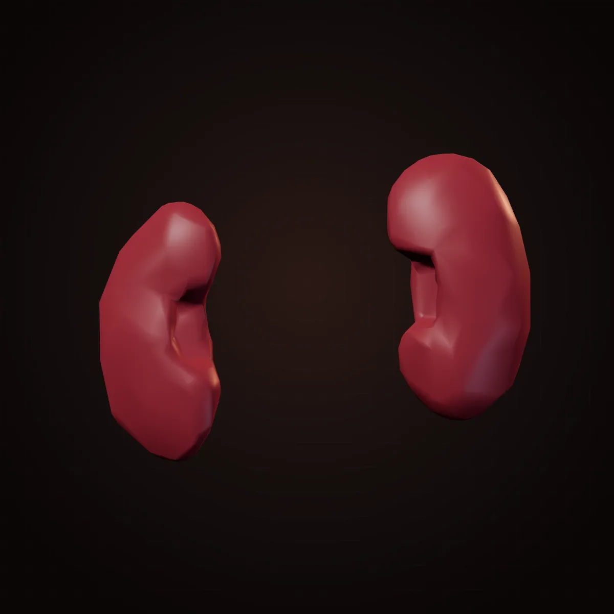
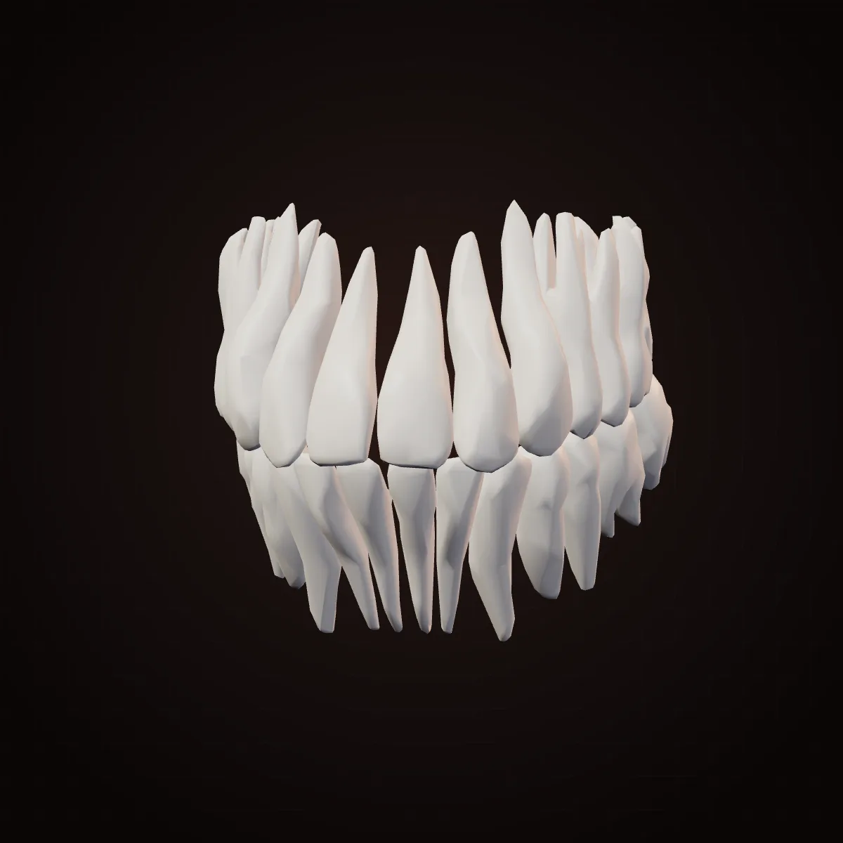
The counts are our own classification, so treat them as a way of seeing rather than a law of nature. But the point stands: a body made of thousands of named structures runs on fewer than twenty geometric ideas.
Protection: domes, cages and cushions
The body protects its most fragile parts with curved shells, flexible cages and fluid. Each handles impact differently, and each is matched to what it guards.
The skull is a dome of eight curved plates. A dome encloses the most volume for the least material and spreads a blow across its whole curve instead of concentrating it at a point. The plates meet in zig-zag seams (sutures) that interlock like a jigsaw. At birth they have not fused, which lets the head mould through the birth canal and the skull keep growing; the soft spot on top usually closes by about 18 to 24 months1.
The rib cage is a set of flexible hoops. Ribs are curved, flat bones that slope downward towards the front. A rigid box would protect the heart and lungs but could not breathe; hoops that swing up and out — like lifting a bucket handle — enlarge the chest with each breath while still absorbing a blow. The lowest two pairs do not reach the breastbone at all, leaving the waist free to bend.
The brain floats. Three membranes wrap it, with cerebrospinal fluid between the inner layers. Buoyancy matters more than most people realise: a brain of about 1.4 kg presses down with the effective weight of roughly 50 g, so it does not crush its own blood vessels and lower structures1.
Transport: tubes, pumps and one-way valves
Anything that has to travel through the body — blood, air, food, urine, lymph — travels in a tube, and almost every tube has a way to keep flow going in one direction.
The heart is two pumps in one cone. The right side pushes blood a short distance through the lungs; the left side pushes it around the whole body, so the left ventricle's wall is much thicker. Its muscle fibres wrap in spirals, so each beat wrings the heart like a towel rather than squeezing it flat. Its four valves are pocket-shaped flaps: backflow fills the pockets and slams them shut, which is the "lub-dub" you hear.
Arteries and veins are built for their pressures. Arteries carry high-pressure blood, so their walls are thick, muscular and elastic; the aorta stretches with each beat and recoils between beats, smoothing the flow. Veins carry low-pressure blood uphill from the legs, so they are thin and floppy, with one-way valves inside. Every step you take squeezes them, and the valves stop the blood sliding back down.
The windpipe is held open by C-shaped rings. The rings keep the airway from collapsing when you breathe in hard, and they are open at the back where the oesophagus sits, so a swallowed mouthful can bulge into that space. The oesophagus itself does not rely on gravity: rings of muscle squeeze food down in waves, which is why astronauts can swallow in orbit.
Even the plumbing has clever valves. Each ureter enters the bladder wall at a slant. As the bladder fills, its own pressure squeezes that tunnel shut — a valve with no moving parts that stops urine flowing back to the kidneys1.
Exchange: why lungs and guts are folded and branched
Organs that exchange gases, nutrients or waste are shaped to maximise surface area, because diffusion only works across very thin barriers over very short distances. Folding and branching are the only practical ways to pack that much surface into a body.
- Air sacs (alveoli) in a pair of adult lungs, on average
- 480 million
- Inner surface of the whole digestive tract, mostly the small intestine
- 32 m²
- Levels of branching from windpipe to air sacs
- 23
- Filtering units (nephrons) in a typical kidney — numbers vary widely
- About 1 million
The lungs are a branching tree ending in grapes. The windpipe splits, and each branch keeps splitting — about 23 times — into ever narrower tubes, delivering air from one pipe to hundreds of millions of tiny sacs. A careful 2004 count found a mean of 480 million alveoli in adult lungs, with a wide range between people that tracked lung size2. Each sac is wrapped in capillaries, and the wall between air and blood is a fraction of a micrometre thick.
The gut multiplies its surface three times over. The small intestine's lining is folded into ridges, the ridges are covered in finger-like villi, and each lining cell carries a brush of microscopic microvilli. Older textbooks claimed the result was the size of a tennis court. A 2014 re-measurement put the inner surface of the entire digestive tract at about 32 square metres — closer to half a badminton court — with roughly 30 m² in the small intestine3. The shape logic is the same; the number was simply wrong for decades.
The brain folds for a different reason. Thinking happens in the cortex, a thin sheet of neurons, so more sheet means more processing. Folding fits a large sheet into a skull small enough to be born. The cerebellum is folded even more finely: it is about a tenth of the brain's mass but holds about 69 of its roughly 86 billion neurons4.
Force: levers, arches and springs
Bones, joints and tendons are shaped to handle force with as little material as possible, using the same three ideas engineers use: levers to multiply movement, arches to carry load, and springs to store and return energy.
Long bones are hollow tubes. Bending loads are carried mostly by the outer wall, so a tube is nearly as strong as a solid rod at a fraction of the weight. Inside the ends, spongy bone forms a lattice of tiny struts aligned along the lines of stress — and bone keeps rebuilding to match its loads, which is why exercise strengthens it and space flight weakens it1.
The kneecap is a pulley. It is the largest sesamoid bone, one that grows inside a tendon. By holding the thigh muscle's tendon away from the knee joint, it lengthens the lever arm, so straightening the knee takes less force.
The foot is a set of springy arches. Its bones form a long arch on the inner side, a lower one on the outside and a cross-wise one in the middle. They flatten slightly as your weight lands and recoil as you push off, storing energy in ligaments and tendons. The Achilles tendon, the thickest in the body, works the same way: it stretches on landing and snaps back on push-off.
The thumb is a saddle. Its base joint is shaped like a horse's saddle, letting the thumb swing in two planes so its pad can meet the pad of each finger. That single movement — opposition — is the basis of both a precision pinch and a power grip.
Sensing: instruments shaped by physics
The sense organs are the clearest proof that shape is function, because each one is shaped by the physics of what it detects.
- The cochlea is a spiral about two and a half turns long. A membrane inside is stiff and narrow at the base, where high pitches vibrate it, and wide and floppy at the tip, where low pitches do. Coiling packs a tube about 3.5 cm long into a space the size of a pea.
- The middle-ear bones are a lever chain. Sound in air would mostly bounce off the fluid of the inner ear. The three ossicles — including the stapes, about 3 mm long and the smallest bone in the body — funnel vibration from the large eardrum onto a tiny footplate, raising the pressure many times over.
- The balance organ is three loops at right angles, one for each direction the head can rotate.
- The eye is a sphere with a dome in front. A sphere rotates freely in its socket while the optics stay aligned; the curved cornea does about two-thirds of the focusing1.
- Eyelashes are a tuned length. A study of 22 mammal species found lashes are typically about one-third of the eye's width — the length that, in wind-tunnel tests, roughly halved both dust reaching the eye and evaporation of tears5.
- The outer ear's folds are a direction finder. Its ridges echo sound slightly differently depending on where it comes from, and the brain uses those tiny differences to tell whether a sound is above or below you.
Could a body part be placed somewhere else?
Some body parts can be moved and keep working — surgeons do it every day — while others only work in one place because their shape depends on their neighbours. The answer depends on what the part needs: a blood supply it can reconnect to, the right temperature, protection, or a precise mechanical fit.
| Part | Where it can go | What happens |
|---|---|---|
| Kidney | Low in the front of the belly | Transplanted kidneys go in the iliac fossa, close to the pelvic vessels and bladder; the old kidneys usually stay |
| Big toe or second toe | The hand | Transferred with its nerves, tendons and vessels to replace a lost thumb |
| Fibula | The jaw | A length of calf bone is shaped into a new lower jaw after cancer surgery |
| Rib cartilage | Under forearm skin, then the head | Carved into an ear and grown in the forearm before attachment (US Army, 2018)7 |
| Parathyroid gland | A forearm muscle | Fragments implanted after neck surgery keep making hormone8 |
| Canine tooth | The eye | Used as a mount for a plastic lens in the "tooth-in-eye" operation9 |
| Donor heart | Beside the patient's own heart | "Piggyback" heterotopic transplants, first done in Cape Town in 1974; rare today10 |
| Scalp hair | Bald areas, eyebrows | Follicles from the back of the head mostly keep their resistance to balding11 |
| Pancreatic islets | The liver | Infused into the portal vein, they lodge in the liver and make insulin |
Table: Body parts that surgeons already move to a different place, and what happens
Some parts move on their own. A kidney sometimes develops in the pelvis instead of the back. The thyroid starts at the back of the tongue in the embryo and descends to the neck; rarely it stays behind. Breast tissue can form anywhere along a "milk line" from the armpit to the groin. Most striking of all, in situs inversus totalis — about 1 in 10,000 people — every organ is a mirror image of the usual layout6.
Some parts tell you why they are where they are. The testes form inside the belly near the kidneys and descend before birth, because sperm production works best at roughly 2–4 °C below core body temperature12. That one requirement explains their unusual position.
Many parts can only be where they are. The cochlea, the hip joint, the heart's valves and the brain all depend on a precise fit with their surroundings. The explorer marks 104 of its 164 parts as "stays put", 43 as already moved by surgeons, 9 as sometimes developing elsewhere on their own and 8 as movable only in part.

Parts that can move
- Have one main artery and vein that can be reconnected (kidney, toe, fibula, muscle flaps)
- Do a job that does not depend on neighbours (hormone glands, islets, fat, skin)
- Are spare or redundant (palmaris longus tendon, a length of fibula, a saphenous vein)
Parts that must stay
- Depend on a precise mechanical fit (joints, ossicles, cochlea, heart valves)
- Need a protected space or short wiring (brain, spinal cord, eyes)
- Are defined by the opening they control (sphincters, larynx, pelvic floor)
Where the design is not perfect
The body is very well adapted, but it is not optimal, because evolution can only modify what already exists. A few famous examples show the history in our anatomy.
- The eye's blind spot. In vertebrates, the retina's wiring lies in front of its light-sensing cells, so the nerve fibres must pass through the retina to leave the eye, creating a spot with no receptors. Octopus eyes, which evolved separately, have the wiring behind and no blind spot.
- The looping laryngeal nerve. The nerve to the voice box runs down into the chest, hooks under a major artery and climbs back up. It made sense in fish ancestors; as the neck lengthened and the heart moved down, the loop stayed. In a giraffe the detour is several metres.
- Wisdom teeth. Human jaws have shortened over our evolution, often leaving no room for the third molars.
- The shared throat. Air and food share a crossroads, guarded by the epiglottis. The human larynx sits low, which helps speech but makes choking easier.
- Leftovers. The coccyx is a tail remnant, goosebumps are a fur reflex, and the palmaris longus tendon is missing in about one in five people with no loss of grip13.
The smallest parts: hair, nails, skin and cells
Even the smallest structures follow the same logic. Nails are rigid backings for the fingertip pad: when you press on something, the pad pushes against the nail, which sharpens touch and grip. Fingerprints are ridges that form in the womb; their exact purpose is still studied, with grip on rough surfaces and finer touch the leading ideas. Hair curls according to the shape of its follicle — round follicles grow straight hair, flattened and curved ones grow curly hair. Skin is thinnest on the eyelids, where movement matters, and thickest on the soles, which take the wear. Neurons are branching trees that collect signals from thousands of others, with one long cable — the motor neurons to your toes run from the lower spine to the foot. Search any of these in the explorer and filter by "Design pattern" to see the same shape idea at every scale.
What designers can learn from the body
The body is the oldest proof that shape communicates function. A handle you grip, a hinge that only bends one way, a valve that closes itself — these are physical affordances, and good interfaces borrow the same idea: the form of a control should tell you what it does. The body also illustrates Gall's law: a complex system that works evolved from a simpler system that worked, which is why the laryngeal nerve still takes its detour. Products accumulate detours the same way. If you are redesigning one, it is worth asking which of your shapes are function and which are history — the same question at the heart of a behavioral UX audit.
How we checked this
Every entry in the guide and the explorer was written against standard anatomy references — principally OpenStax's open textbook Anatomy and Physiology 2e1 — and, for numbers and surprising claims, against the peer-reviewed papers and official reports in the sources. "Could it be somewhere else?" verdicts describe only established surgical practice or documented anatomical variants. Where science is still debated (the role of the sinuses, fingerprints or the appendix), we say so. Counts such as nephrons and alveoli vary a great deal between people, so we give typical values. This is an educational guide, not medical advice. Last reviewed October 2026.
Key terms, defined
- Form follows function — the principle that a structure's shape is explained by the job it does.
- Alveoli — the tiny air sacs at the ends of the airways where oxygen enters the blood.
- Villi — finger-like projections of the intestinal lining that increase its surface area.
- Sesamoid bone — a bone that grows inside a tendon, such as the kneecap.
- Heterotopic transplant — placing a donor organ in a different position from the original one.
- Situs inversus — a mirror-image arrangement of the internal organs.
- Vestigial structure — a reduced part that matches a fully working structure in related animals.
Frequently asked questions
Why is the human body shaped the way it is?
Because each part's shape solves a physical problem. Domes and curves protect, tubes move fluids and food, folds and branching trees create the large surfaces needed to exchange gases and nutrients, and levers and arches handle force. Evolution kept shapes that worked well enough for survival, which is why the same few geometries appear all over the body.
Why are red blood cells shaped like discs?
A red blood cell is a flexible disc thinner in the middle than at the rim. That shape gives a large surface for oxygen to cross and keeps every part of the cell close to the surface. It is also very deformable, so cells fold to squeeze through capillaries narrower than themselves. In sickle cell disease the cells become rigid and curved and get stuck.
Why are the lungs and intestines so folded?
Exchange happens across surfaces, so more surface means more exchange. The lungs end in about 480 million tiny air sacs, and the intestine's lining is folded into villi and microvilli. A 2014 study estimated the inner surface of the whole digestive tract at about 32 square metres, most of it in the small intestine.
Can an organ be moved to a different place in the body?
Some can. Transplanted kidneys are placed low in the front of the belly rather than in the back. Transplanted pancreases go in the lower abdomen too. Parathyroid tissue can be implanted in a forearm muscle, a big toe can replace a thumb, and an ear can be grown under forearm skin before being attached. Others, such as the brain or cochlea, only work where they are.
Can a person have their organs on the wrong side?
Yes. In situs inversus totalis, about 1 in 10,000 people, all the organs are a mirror image of the usual layout: the heart points right, the liver is on the left. Many people with it are healthy and only find out through a scan for something else, though it can come with other conditions.
Is the human body perfectly designed?
No. It is very well adapted, but evolution modifies what already exists rather than starting fresh. The eye's wiring runs in front of its light sensors, creating a blind spot; the nerve to the voice box loops down into the chest and back; and shorter jaws leave many people without room for wisdom teeth.
Which body parts can you live without?
People live normally without their appendix, gallbladder, spleen (with extra vaccinations), tonsils, one kidney, and a length of fibula. Some can even live without a stomach by eating small, frequent meals. Each loss has some cost, so removal is only done for a medical reason.
Sources
- OpenStax — Anatomy and Physiology 2e, Rice University (2022)
- Ochs et al. — The number of alveoli in the human lung, Am J Respir Crit Care Med (2004)
- Helander & Fändriks — Surface area of the digestive tract, revisited, Scand J Gastroenterol (2014)
- Azevedo et al. — Equal numbers of neuronal and nonneuronal cells make the human brain an isometrically scaled-up primate brain, J Comp Neurol (2009)
- Amador et al. — Eyelashes divert airflow to protect the eye, J R Soc Interface (2015)
- Radiopaedia — Situs inversus
- US Army — Army surgeon transplants ear 'grown' on Soldier's forearm (2018)
- ClinicalTrials.gov — Parathyroid auto-transplantation: randomized trial of the personalized site (NCT02906748)
- Journal of Indian Prosthodontic Society — review of osteo-odonto-keratoprosthesis (2018)
- PubMed Central — Heterotopic heart transplant history and concepts cannot be neglected
- Orentreich — Autografts in alopecias and other selected dermatological conditions, Ann N Y Acad Sci (1959)
- Frontiers in Cell and Developmental Biology — review of heat stress and testicular function (2022)
- Yammine — Clinical prevalence of palmaris longus agenesis: a systematic review and meta-analysis, Clin Anat (2013)
- BodyParts3D, © The Database Center for Life Science, licensed under CC Attribution 4.0 International
- Checked October 25, 2026. Figures that change — fees, salaries, prices — are reviewed every six months; if something is out of date, tell us.
Where to go from here
- Human body explorer — rotate a real 3D scan of the body and click any of 164 parts
- Affordance — why a shape tells you how to use it, in products as in bodies
- Everyday design scienceWhy are stop signs octagons? The design rules behind road signs around the worldStop signs are octagons so drivers recognise them by shape alone, even from behind. The history of road sign shapes, colours and lettering from 1923 to today.
- Everyday design scienceLondon Tube map history: why Harry Beck's 1933 diagram isn't to scale, and why that worksLondon Tube map history from the 1908 geographic maps to Harry Beck's 1933 diagram. Why it isn't to scale, how it shapes route choices and what research shows.
- Everyday design scienceWhy are traffic lights red, yellow and green? The railway history and the science of seeing themTraffic lights are red, yellow and green because road signals copied railway signals. The history, the physics of each colour and how colour-blind drivers cope.
- How to make an appWhat is a PWA? Progressive web apps explained simplyA PWA is a website that installs and works like an app, offline and with push. How PWAs work, Indian success stories, iPhone limits and when to choose one.
- Tech stackWhat is an API? A plain-English explanation with a restaurant analogy and real app examplesWhat is an API? A plain-English explanation using a restaurant analogy, with real examples from UPI, maps and login, plus REST, keys, webhooks and security.
- Tech explainedHow does ChatGPT work? Tokens, transformers, training and why it sometimes makes things upHow ChatGPT works, in plain English: it predicts the next token using a transformer trained on vast text, then tuned with human feedback. With diagrams.
Get the ideas, not the noise.
Sign up with Google and we’ll send you short UX psychology tips by email — why people tap, stall and leave, and what to do about it. Unsubscribe anytime. Prefer something to keep? Get the free Laws of UX guide as a PDF.
The same science shapes your product.
A behavioral UX audit of your app or SaaS product: why users hesitate, stall and leave, and a prioritized fix list. Fixed price, 2–4 weeks.